[English] 日本語
Yorodumi
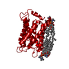
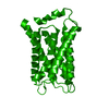
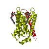
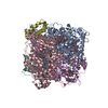
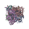
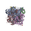
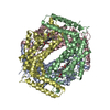
- PDB-2b6p: X-ray structure of lens Aquaporin-0 (AQP0) (lens MIP) in an open ... -

+
Open data


ID or keywords:

Loading...

-
Basic information

Entry
Database: PDB / ID: 2b6p
TitleX-ray structure of lens Aquaporin-0 (AQP0) (lens MIP) in an open pore state
ComponentsLens fiber major intrinsic protein
KeywordsMEMBRANE PROTEIN / aquaporin-0 / AQP0 / lens MIP / open water pore / aquaporin
Function / homology
Function and homology information


Passive transport by Aquaporins / gap junction-mediated intercellular transport / water channel activity / water transport / structural constituent of eye lens / gap junction / lens development in camera-type eye / positive regulation of cell adhesion / visual perception / protein homotetramerization ...Passive transport by Aquaporins / gap junction-mediated intercellular transport / water channel activity / water transport / structural constituent of eye lens / gap junction / lens development in camera-type eye / positive regulation of cell adhesion / visual perception / protein homotetramerization / calmodulin binding / apical plasma membrane / endoplasmic reticulum / plasma membrane
Similarity search - Function
Glycerol uptake facilitator protein / Glycerol uptake facilitator protein. / Aquaporin transporter / Major intrinsic protein, conserved site / MIP family signature. / Major intrinsic protein / Major intrinsic protein / Aquaporin-like / Up-down Bundle / Mainly Alpha
Similarity search - Domain/homology
Lens fiber major intrinsic protein
Similarity search - Component
Biological speciesBos taurus (cattle)
MethodX-RAY DIFFRACTION / MOLECULAR REPLACEMENT / Resolution: 2.4 Å
AuthorsGonen, T. / Cheng, Y. / Sliz, P. / Hiroaki, Y. / Fujiyoshi, Y. / Harrison, S.C. / Walz, T.
CitationJournal: Proc.Natl.Acad.Sci.USA / Year: 2004
Title: The Channel Architecture Of Aquaporin O At 2.2 Angstrom Resolution
Authors: Harries, W.E.C. / Akhavan, D. / Miercke, L.J.W. / Khademi, S. / Stroud, R.M.
History
DepositionOct 3, 2005Deposition site: RCSB / Processing site: RCSB
Revision 1.0Dec 6, 2005Provider: repository / Type: Initial release
Revision 1.1May 1, 2008Group: Version format compliance
Revision 1.2Jul 13, 2011Group: Version format compliance
Revision 1.3Oct 26, 2011Group: Other
Revision 1.4Feb 14, 2024Group: Data collection / Database references / Category: chem_comp_atom / chem_comp_bond / database_2
Item: _database_2.pdbx_DOI / _database_2.pdbx_database_accession
Remark 0THIS ENTRY 2B6P REFLECTS AN ALTERNATIVE MODELING OF THE STRUCTURAL DATA IN R1YMGSF ORIGINAL DATA ...THIS ENTRY 2B6P REFLECTS AN ALTERNATIVE MODELING OF THE STRUCTURAL DATA IN R1YMGSF ORIGINAL DATA DETERMINED BY AUTHOR: W.E.C.HARRIES,D.AKHAVAN,L.J.W.MIERCKE,S.KHADEMI, R.M.STROUD.

-
Structure visualization

Structure viewerMolecule:
MolmilJmol/JSmol

Downloads & links

-
Assembly

Deposited unit
A: Lens fiber major intrinsic protein


Theoretical massNumber of molelcules
Total (without water)28,2451
Polymers28,2451
Non-polymers00
Water2,324129
1
A: Lens fiber major intrinsic protein

A: Lens fiber major intrinsic protein

A: Lens fiber major intrinsic protein

A: Lens fiber major intrinsic protein


Theoretical massNumber of molelcules
Total (without water)112,9794
Polymers112,9794
Non-polymers00
Water724
TypeNameSymmetry operationNumber
identity operation1_555x,y,z1
crystal symmetry operation2_565-x,-y+1,z1
crystal symmetry operation3_555-y+1/2,x+1/2,z1
crystal symmetry operation4_455y-1/2,-x+1/2,z1
Buried area13830 Å2
ΔGint-125 kcal/mol
Surface area40440 Å2
MethodPISA
Unit cell
Length a, b, c (Å)109.531, 109.531, 52.822
Angle α, β, γ (deg.)90.00, 90.00, 90.00
Int Tables number90
Space group name H-MP4212

-
Components

#1: Protein Lens fiber major intrinsic protein / / Aquaporin-0 / MIP26 / MP26


Mass: 28244.865 Da / Num. of mol.: 1 / Source method: isolated from a natural source / Source: (natural) Bos taurus (cattle) / References: UniProt: P06624
#2: Water ChemComp-HOH / water / Water


Mass: 18.015 Da / Num. of mol.: 129 / Source method: isolated from a natural source / Formula: H2O

-
Experimental details

-
Experiment

ExperimentMethod: X-RAY DIFFRACTION

-
Sample preparation

CrystalDensity Matthews: 2.8 Å3/Da / Density % sol: 56.14 % / Description: AUTHOR USED THE SF DATA FROM ENTRY 1YMG.
Crystal growTemperature: 300 K / Method: vapor diffusion, sitting drop / pH: 10
Details: 20 mM glycine, pH 10.0 30% polyethylene glycol 1K 50 mM NaCl, VAPOR DIFFUSION, SITTING DROP, temperature 300K

-
Data collection

RadiationProtocol: SINGLE WAVELENGTH / Monochromatic (M) / Laue (L): M / Scattering type: x-ray
Radiation wavelengthRelative weight: 1

-
Processing

SoftwareName: CNS / Version: 1.1 / Classification: refinement
RefinementMethod to determine structure: MOLECULAR REPLACEMENT / Resolution: 2.4→14.88 Å / Rfactor Rfree error: 0.008 / Data cutoff high absF: 344976.22 / Data cutoff low absF: 0 / Isotropic thermal model: RESTRAINED / Cross valid method: THROUGHOUT / σ(F): 0 / Stereochemistry target values: Engh & Huber
RfactorNum. reflection% reflectionSelection details
Rfree0.292 1218 10.1 %RANDOM
Rwork0.242 ---
all-0 --
obs-12037 92.6 %-
Displacement parametersBiso mean: 63.2 Å2
Baniso -1Baniso -2Baniso -3
1--11.45 Å20 Å20 Å2
2---11.45 Å20 Å2
3---22.91 Å2
Refine analyze
FreeObs
Luzzati coordinate error0.37 Å0.29 Å
Luzzati d res low-5 Å
Luzzati sigma a0.33 Å0.23 Å
Refinement stepCycle: LAST / Resolution: 2.4→14.88 Å
ProteinNucleic acidLigandSolventTotal
Num. atoms1982 0 0 129 2111
Refine LS restraints
Refine-IDTypeDev ideal
X-RAY DIFFRACTIONo_bond_d0.006
X-RAY DIFFRACTIONo_angle_deg1.3
X-RAY DIFFRACTIONo_dihedral_angle_d19.8
X-RAY DIFFRACTIONo_improper_angle_d0.9
LS refinement shellResolution: 2.4→2.55 Å / Rfactor Rfree error: 0.024 / Total num. of bins used: 6
RfactorNum. reflection% reflection
Rfree0.331 185 10.5 %
Rwork0.265 1581 -
obs--84.1 %
Xplor file
Refine-IDSerial noParam fileTopol file
X-RAY DIFFRACTION1protein_rep.paramprotein.top
X-RAY DIFFRACTION2water.param

+
About Yorodumi

-
News

-
Feb 9, 2022. New format data for meta-information of EMDB entries

New format data for meta-information of EMDB entries

  • Version 3 of the EMDB header file is now the official format.
  • The previous official version 1.9 will be removed from the archive.

Related info.:EMDB header

External links:wwPDB to switch to version 3 of the EMDB data model

-
Aug 12, 2020. Covid-19 info

Covid-19 info

URL: https://pdbj.org/emnavi/covid19.php

New page: Covid-19 featured information page in EM Navigator.

Related info.:Covid-19 info / Mar 5, 2020. Novel coronavirus structure data

+
Mar 5, 2020. Novel coronavirus structure data

Novel coronavirus structure data

Related info.:Yorodumi Speices / Aug 12, 2020. Covid-19 info

External links:COVID-19 featured content - PDBj / Molecule of the Month (242):Coronavirus Proteases

+
Jan 31, 2019. EMDB accession codes are about to change! (news from PDBe EMDB page)

EMDB accession codes are about to change! (news from PDBe EMDB page)

  • The allocation of 4 digits for EMDB accession codes will soon come to an end. Whilst these codes will remain in use, new EMDB accession codes will include an additional digit and will expand incrementally as the available range of codes is exhausted. The current 4-digit format prefixed with “EMD-” (i.e. EMD-XXXX) will advance to a 5-digit format (i.e. EMD-XXXXX), and so on. It is currently estimated that the 4-digit codes will be depleted around Spring 2019, at which point the 5-digit format will come into force.
  • The EM Navigator/Yorodumi systems omit the EMD- prefix.

Related info.:Q: What is EMD? / ID/Accession-code notation in Yorodumi/EM Navigator

External links:EMDB Accession Codes are Changing Soon! / Contact to PDBj

+
Jul 12, 2017. Major update of PDB

Major update of PDB

  • wwPDB released updated PDB data conforming to the new PDBx/mmCIF dictionary.
  • This is a major update changing the version number from 4 to 5, and with Remediation, in which all the entries are updated.
  • In this update, many items about electron microscopy experimental information are reorganized (e.g. em_software).
  • Now, EM Navigator and Yorodumi are based on the updated data.

External links:wwPDB Remediation / Enriched Model Files Conforming to OneDep Data Standards Now Available in the PDB FTP Archive

-
Yorodumi

Thousand views of thousand structures

  • Yorodumi is a browser for structure data from EMDB, PDB, SASBDB, etc.
  • This page is also the successor to EM Navigator detail page, and also detail information page/front-end page for Omokage search.
  • The word "yorodu" (or yorozu) is an old Japanese word meaning "ten thousand". "mi" (miru) is to see.

Related info.:EMDB / PDB / SASBDB / Comparison of 3 databanks / Yorodumi Search / Aug 31, 2016. New EM Navigator & Yorodumi / Yorodumi Papers / Jmol/JSmol / Function and homology information / Changes in new EM Navigator and Yorodumi

Read more