 Movie
Movie Controller

[English] 日本語
 Yorodumi
- PDB-5l6v: Crystal structure of E. coli ADP-glucose pyrophosphorylase (AGPas... -
+ Open data

- Basic information
| Entry | Database: PDB / ID: 5l6v | |||||||||
|---|---|---|---|---|---|---|---|---|---|---|
| Title | Crystal structure of E. coli ADP-glucose pyrophosphorylase (AGPase) in complex with a negative allosteric regulator adenosine monophosphate (AMP) - AGPase*AMP | |||||||||
 Components Components | Glucose-1-phosphate adenylyltransferase | |||||||||
 Keywords Keywords | TRANSFERASE | |||||||||
| Function / homology |  Function and homology information Function and homology informationglucose-1-phosphate adenylyltransferase complex / glucose-1-phosphate adenylyltransferase / glucose-1-phosphate adenylyltransferase activity / glucan biosynthetic process / glycogen biosynthetic process / AMP binding / protein homotetramerization / magnesium ion binding / ATP binding / identical protein binding Similarity search - Function | |||||||||
| Biological species |  | |||||||||
| Method |  X-RAY DIFFRACTION / SYNCHROTRON / MOLECULAR REPLACEMENT / Resolution: 2.667 Å X-RAY DIFFRACTION / SYNCHROTRON / MOLECULAR REPLACEMENT / Resolution: 2.667 Å | |||||||||
 Authors Authors | Cifuente, J.O. / Albesa-Jove, D. / Comino, N. / Madariaga-Marcos, J. / Agirre, J. / Lopez-Fernandez, S. / Garcia-Alija, M. / Guerin, M.E. | |||||||||
 Citation Citation | Journal: Structure / Year: 2016 Title: Structural Basis of Glycogen Biosynthesis Regulation in Bacteria. Authors: Cifuente, J.O. / Comino, N. / Madariaga-Marcos, J. / Lopez-Fernandez, S. / Garcia-Alija, M. / Agirre, J. / Albesa-Jove, D. / Guerin, M.E. | |||||||||
| History |
|
- Structure visualization
| Structure viewer | Molecule: MolmilJmol/JSmol |
|---|
- Downloads & links
-Download
| PDBx/mmCIF format | 5l6v.cif.gz | 1.2 MB | Display | PDBx/mmCIF format |
|---|---|---|---|---|
| PDB format | pdb5l6v.ent.gz | 1 MB | Display | PDB format |
| PDBx/mmJSON format | 5l6v.json.gz | Tree view | PDBx/mmJSON format | |
| Others |  Other downloads Other downloads |
-Validation report
| Arichive directory | https://data.pdbj.org/pub/pdb/validation_reports/l6/5l6vftp://data.pdbj.org/pub/pdb/validation_reports/l6/5l6v | HTTPS FTP |
|---|
-Related structure data
| Related structure data |  5l6sC  1yp2S S: Starting model for refinement C: citing same article ( |
|---|---|
| Similar structure data |


-Links
 PDBj
PDBj


- Assembly
| Deposited unit | 
| ||||||||
|---|---|---|---|---|---|---|---|---|---|
| 1 | 
| ||||||||
| 2 | 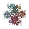
| ||||||||
| 3 | 
| ||||||||
| 4 | 
| ||||||||
| Unit cell |
|
-Components
| #1: Protein | Mass: 48758.590 Da / Num. of mol.: 16 Source method: isolated from a genetically manipulated source Source: (gene. exp.) References: UniProt: P0A6V1, glucose-1-phosphate adenylyltransferase #2: Polysaccharide | beta-D-fructofuranose-(2-1)-alpha-D-glucopyranose / sucrose   Source method: isolated from a genetically manipulated source Details: oligosaccharide with reducing-end-to-reducing-end glycosidic bond References: sucrose #3: Chemical | ChemComp-AMP /   Mass: 347.221 Da / Num. of mol.: 16 / Source method: obtained synthetically / Formula: C10H14N5O7P / Comment: AMP*YM Mass: 347.221 Da / Num. of mol.: 16 / Source method: obtained synthetically / Formula: C10H14N5O7P / Comment: AMP*YM#4: Chemical | ChemComp-PO4 / |   Mass: 94.971 Da / Num. of mol.: 1 / Source method: obtained synthetically / Formula: PO4 Mass: 94.971 Da / Num. of mol.: 1 / Source method: obtained synthetically / Formula: PO4#5: Water | ChemComp-HOH / |  Mass: 18.015 Da / Num. of mol.: 1398 / Source method: isolated from a natural source / Formula: H2O Mass: 18.015 Da / Num. of mol.: 1398 / Source method: isolated from a natural source / Formula: H2O |
|---|
-Experimental details
-Experiment
| Experiment | Method: X-RAY DIFFRACTION / Number of used crystals: 1 |
|---|
- Sample preparation
| Crystal | Density Matthews: 2.58 Å3/Da / Density % sol: 52.29 % |
|---|---|
| Crystal grow | Temperature: 291 K / Method: vapor diffusion, sitting drop / pH: 7 Details: 10mM EDTA, 10% D-(+)-Sucrose, 50mM MgCl2, 5mM ADP, 16% (w/v) PEG 10000, 100mM Imidazole pH 7.00 |
-Data collection
| Diffraction | Mean temperature: 100 K |
|---|---|
| Diffraction source | Source: SYNCHROTRON / Site: Diamond  / Beamline: I04 / Wavelength: 0.97625 Å / Beamline: I04 / Wavelength: 0.97625 Å |
| Detector | Type: DECTRIS PILATUS 6M-F / Detector: PIXEL / Date: May 23, 2013 |
| Radiation | Protocol: SINGLE WAVELENGTH / Monochromatic (M) / Laue (L): M / Scattering type: x-ray |
| Radiation wavelength | Wavelength: 0.97625 Å / Relative weight: 1 |
| Reflection | Resolution: 2.667→45.271 Å / Num. obs: 224803 / % possible obs: 100 % / Redundancy: 3.2 % / Biso Wilson estimate: 30.64 Å2 / CC1/2: 0.959 / Rmerge(I) obs: 0.2 / Net I/σ(I): 5.69 |
| Reflection shell | Resolution: 2.667→2.762 Å / Redundancy: 3.2 % / Rmerge(I) obs: 0.7832 / Mean I/σ(I) obs: 1.74 / % possible all: 100 |
- Processing
| Software |
| |||||||||||||||||||||||||||||||||||||||||||||||||||||||||||||||||||||||||||||||||||||||||||||||||||||||||||||||||||||||||||||||||||||||||||||||||||||||||||||||||||||||||||||||||||||||||||||||||||||||||||||||||||||||||
|---|---|---|---|---|---|---|---|---|---|---|---|---|---|---|---|---|---|---|---|---|---|---|---|---|---|---|---|---|---|---|---|---|---|---|---|---|---|---|---|---|---|---|---|---|---|---|---|---|---|---|---|---|---|---|---|---|---|---|---|---|---|---|---|---|---|---|---|---|---|---|---|---|---|---|---|---|---|---|---|---|---|---|---|---|---|---|---|---|---|---|---|---|---|---|---|---|---|---|---|---|---|---|---|---|---|---|---|---|---|---|---|---|---|---|---|---|---|---|---|---|---|---|---|---|---|---|---|---|---|---|---|---|---|---|---|---|---|---|---|---|---|---|---|---|---|---|---|---|---|---|---|---|---|---|---|---|---|---|---|---|---|---|---|---|---|---|---|---|---|---|---|---|---|---|---|---|---|---|---|---|---|---|---|---|---|---|---|---|---|---|---|---|---|---|---|---|---|---|---|---|---|---|---|---|---|---|---|---|---|---|---|---|---|---|---|---|---|---|
| Refinement | Method to determine structure: MOLECULAR REPLACEMENT Starting model: 1YP2 Resolution: 2.667→45.271 Å / SU ML: 0.42 / Cross valid method: FREE R-VALUE / σ(F): 0.44 / Phase error: 28.2 / Stereochemistry target values: ML
| |||||||||||||||||||||||||||||||||||||||||||||||||||||||||||||||||||||||||||||||||||||||||||||||||||||||||||||||||||||||||||||||||||||||||||||||||||||||||||||||||||||||||||||||||||||||||||||||||||||||||||||||||||||||||
| Solvent computation | Shrinkage radii: 0.9 Å / VDW probe radii: 1.11 Å / Solvent model: FLAT BULK SOLVENT MODEL | |||||||||||||||||||||||||||||||||||||||||||||||||||||||||||||||||||||||||||||||||||||||||||||||||||||||||||||||||||||||||||||||||||||||||||||||||||||||||||||||||||||||||||||||||||||||||||||||||||||||||||||||||||||||||
| Refinement step | Cycle: LAST / Resolution: 2.667→45.271 Å
| |||||||||||||||||||||||||||||||||||||||||||||||||||||||||||||||||||||||||||||||||||||||||||||||||||||||||||||||||||||||||||||||||||||||||||||||||||||||||||||||||||||||||||||||||||||||||||||||||||||||||||||||||||||||||
| Refine LS restraints |
| |||||||||||||||||||||||||||||||||||||||||||||||||||||||||||||||||||||||||||||||||||||||||||||||||||||||||||||||||||||||||||||||||||||||||||||||||||||||||||||||||||||||||||||||||||||||||||||||||||||||||||||||||||||||||
| LS refinement shell |
|
