 Movie
Movie Controller
 Structure viewers
 About Yorodumi Papers


+Search query
-Structure paper
| Title | Lipid-protein interactions in double-layered two-dimensional AQP0 crystals. |
|---|---|
| Journal, issue, pages | Nature, Vol. 438, Issue 7068, Page 633-638, Year 2005 |
| Publish date | Dec 1, 2005 |
 Authors Authors | Tamir Gonen / Yifan Cheng / Piotr Sliz / Yoko Hiroaki / Yoshinori Fujiyoshi / Stephen C Harrison / Thomas Walz /  |
| PubMed Abstract | Lens-specific aquaporin-0 (AQP0) functions as a specific water pore and forms the thin junctions between fibre cells. Here we describe a 1.9 A resolution structure of junctional AQP0, determined by ...Lens-specific aquaporin-0 (AQP0) functions as a specific water pore and forms the thin junctions between fibre cells. Here we describe a 1.9 A resolution structure of junctional AQP0, determined by electron crystallography of double-layered two-dimensional crystals. Comparison of junctional and non-junctional AQP0 structures shows that junction formation depends on a conformational switch in an extracellular loop, which may result from cleavage of the cytoplasmic amino and carboxy termini. In the centre of the water pathway, the closed pore in junctional AQP0 retains only three water molecules, which are too widely spaced to form hydrogen bonds with each other. Packing interactions between AQP0 tetramers in the crystalline array are mediated by lipid molecules, which assume preferred conformations. We were therefore able to build an atomic model for the lipid bilayer surrounding the AQP0 tetramers, and we describe lipid-protein interactions. |
 External links External links | Nature / PubMed:16319884 / PubMed Central |
| Methods | EM (electron crystallography) / X-ray diffraction |
| Resolution | 1.9 - 2.4 Å |
| Structure data | 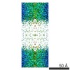 EMDB-2973:  PDB-2b6o: 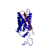 PDB-2b6p: |
| Chemicals |  ChemComp-MC3:  ChemComp-HOH: |
| Source |
|
 Keywords Keywords | MEMBRANE PROTEIN / aquaporin-0 junctions / AQP0 / lens MIP / lipid-protein interactions / membrane / lipid bilayer / closed water pore / aquaporin-0; AQP0; lens MIP; open water pore; aquaporin; membrane protein; |

