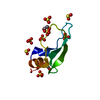
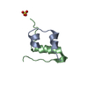
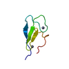
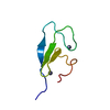
Mass: 8340.073 Da / Num. of mol.: 1 / Mutation: C17U, A18C / Source method: obtained synthetically Details: Chemically synthesized oligonucleotide (RNA). This sequence is a mutated form of that naturally found in Potato leafroll virus References: GenBank: 23953793
Resolution: 1.34→23.6 Å / Cross valid method: THROUGHOUT / σ(F): 4 Stereochemistry target values: G. Parkinson et. al., Acta Cryst. (1996) D52, 57-64. Details: The waters 603, 686, 211, 584, 610, 666, 608 and 638 have partial occupancies. Thus close contacts between them should be ignored.
Rfactor
Num. reflection
% reflection
Selection details
Rwork
0.114
-
-
-
obs
-
18322
89 %
-
Rfree
-
-
-
Random
Refinement step
Cycle: LAST / Resolution: 1.34→23.6 Å
Protein
Nucleic acid
Ligand
Solvent
Total
Num. atoms
0
552
2
201
755
Refine LS restraints
Refine-ID
Type
Dev ideal
X-RAY DIFFRACTION
s_bond_d
0.022
X-RAY DIFFRACTION
s_angle_d
0.041
+
About Yorodumi
-
News
-
Feb 9, 2022. New format data for meta-information of EMDB entries
New format data for meta-information of EMDB entries
Version 3 of the EMDB header file is now the official format.
The previous official version 1.9 will be removed from the archive.
In the structure databanks used in Yorodumi, some data are registered as the other names, "COVID-19 virus" and "2019-nCoV". Here are the details of the virus and the list of structure data.
Jan 31, 2019. EMDB accession codes are about to change! (news from PDBe EMDB page)
EMDB accession codes are about to change! (news from PDBe EMDB page)
The allocation of 4 digits for EMDB accession codes will soon come to an end. Whilst these codes will remain in use, new EMDB accession codes will include an additional digit and will expand incrementally as the available range of codes is exhausted. The current 4-digit format prefixed with “EMD-” (i.e. EMD-XXXX) will advance to a 5-digit format (i.e. EMD-XXXXX), and so on. It is currently estimated that the 4-digit codes will be depleted around Spring 2019, at which point the 5-digit format will come into force.
The EM Navigator/Yorodumi systems omit the EMD- prefix.
Related info.:Q: What is EMD? / ID/Accession-code notation in Yorodumi/EM Navigator
Yorodumi is a browser for structure data from EMDB, PDB, SASBDB, etc.
This page is also the successor to EM Navigator detail page, and also detail information page/front-end page for Omokage search.
The word "yorodu" (or yorozu) is an old Japanese word meaning "ten thousand". "mi" (miru) is to see.
Related info.:EMDB / PDB / SASBDB / Comparison of 3 databanks / Yorodumi Search / Aug 31, 2016. New EM Navigator & Yorodumi / Yorodumi Papers / Jmol/JSmol / Function and homology information / Changes in new EM Navigator and Yorodumi
 Movie
Movie Controller

 Yorodumi
 Open data
 Basic information
 Components
 Keywords
 Function and homology information
 X-RAY DIFFRACTION /
  Authors
 Citation
 Structure visualization
 Downloads & links
 Other downloads


 PDBj
PDBj


 Assembly

 Mass: 24.305 Da / Num. of mol.: 2 / Source method: obtained synthetically / Formula: Mg
 Mass: 18.015 Da / Num. of mol.: 201 / Source method: isolated from a natural source / Formula: H2O
 Sample preparation
 / Beamline: 5ID-B / Wavelength: 0.9198 Å
 Processing