 Movie
Movie Controller

[English] 日本語
 Yorodumi
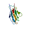
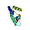
- PDB-6dmq: Crystal structure of the T27A mutant of human alpha defensin HNP4. -
+ Open data

- Basic information
| Entry | Database: PDB / ID: 6dmq | ||||||
|---|---|---|---|---|---|---|---|
| Title | Crystal structure of the T27A mutant of human alpha defensin HNP4. | ||||||
 Components Components | Neutrophil defensin 4 | ||||||
 Keywords Keywords | ANTIMICROBIAL PROTEIN / HUMAN ALPHA-DEFENSIN / ANTIMICROBIAL PEPTIDE | ||||||
| Function / homology |  Function and homology information Function and homology informationdisruption of plasma membrane integrity in another organism / Defensins / pore-forming activity / antifungal humoral response / antimicrobial humoral response / Alpha-defensins / azurophil granule / defense response to fungus / transport vesicle / Golgi lumen ...disruption of plasma membrane integrity in another organism / Defensins / pore-forming activity / antifungal humoral response / antimicrobial humoral response / Alpha-defensins / azurophil granule / defense response to fungus / transport vesicle / Golgi lumen / specific granule lumen / antimicrobial humoral immune response mediated by antimicrobial peptide / antibacterial humoral response / killing of cells of another organism / defense response to Gram-negative bacterium / defense response to Gram-positive bacterium / innate immune response / Neutrophil degranulation / protein homodimerization activity / : / extracellular region Similarity search - Function | ||||||
| Biological species |  Homo sapiens (human) Homo sapiens (human) | ||||||
| Method |  X-RAY DIFFRACTION / SYNCHROTRON / MOLECULAR REPLACEMENT / Resolution: 1.7 Å X-RAY DIFFRACTION / SYNCHROTRON / MOLECULAR REPLACEMENT / Resolution: 1.7 Å | ||||||
 Authors Authors | Gohain, N. / Tolbert, W.D. / Pazgier, M. | ||||||
 Citation Citation | Journal: Biochim Biophys Acta Biomembr / Year: 2019 Title: Systematic mutational analysis of human neutrophil alpha-defensin HNP4. Authors: Hu, H. / Di, B. / Tolbert, W.D. / Gohain, N. / Yuan, W. / Gao, P. / Ma, B. / He, Q. / Pazgier, M. / Zhao, L. / Lu, W. | ||||||
| History |
|
- Structure visualization
| Structure viewer | Molecule: MolmilJmol/JSmol |
|---|
- Downloads & links
-Download
| PDBx/mmCIF format | 6dmq.cif.gz | 112.8 KB | Display | PDBx/mmCIF format |
|---|---|---|---|---|
| PDB format | pdb6dmq.ent.gz | 90.7 KB | Display | PDB format |
| PDBx/mmJSON format | 6dmq.json.gz | Tree view | PDBx/mmJSON format | |
| Others |  Other downloads Other downloads |
-Validation report
| Arichive directory | https://data.pdbj.org/pub/pdb/validation_reports/dm/6dmqftp://data.pdbj.org/pub/pdb/validation_reports/dm/6dmq | HTTPS FTP |
|---|
-Related structure data
| Related structure data |  6dmmC  1zmmS S: Starting model for refinement C: citing same article ( |
|---|---|
| Similar structure data |


-Links
 PDBj
PDBj


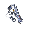
- Assembly
| Deposited unit | 
| ||||||||
|---|---|---|---|---|---|---|---|---|---|
| 1 | 
| ||||||||
| 2 | 
| ||||||||
| 3 | 
| ||||||||
| 4 | 
| ||||||||
| Unit cell |
|
-Components
| #1: Protein/peptide | Mass: 3690.460 Da / Num. of mol.: 8 / Mutation: T27A / Source method: obtained synthetically / Source: (synth.) Homo sapiens (human) / References: UniProt: P12838#2: Chemical | ChemComp-MPD / ( |   Mass: 118.174 Da / Num. of mol.: 1 / Source method: obtained synthetically / Formula: C6H14O2 / Comment: precipitant*YM Mass: 118.174 Da / Num. of mol.: 1 / Source method: obtained synthetically / Formula: C6H14O2 / Comment: precipitant*YM#3: Water | ChemComp-HOH / |  Mass: 18.015 Da / Num. of mol.: 172 / Source method: isolated from a natural source / Formula: H2O Mass: 18.015 Da / Num. of mol.: 172 / Source method: isolated from a natural source / Formula: H2OHas protein modification | Y | |
|---|
-Experimental details
-Experiment
| Experiment | Method: X-RAY DIFFRACTION / Number of used crystals: 1 |
|---|
- Sample preparation
| Crystal | Density Matthews: 2.12 Å3/Da / Density % sol: 41.96 % |
|---|---|
| Crystal grow | Temperature: 294 K / Method: vapor diffusion, sitting drop / pH: 5.5 Details: 30% MPD 15% PEG 8000 100 mM sodium acetate pH 5.5 100 mM calcium chloride |
-Data collection
| Diffraction | Mean temperature: 100 K |
|---|---|
| Diffraction source | Source: SYNCHROTRON / Site: SSRL  / Beamline: BL14-1 / Wavelength: 0.97946 Å / Beamline: BL14-1 / Wavelength: 0.97946 Å |
| Detector | Type: MARMOSAIC 325 mm CCD / Detector: CCD / Date: Jan 7, 2016 |
| Radiation | Monochromator: Si (111) / Protocol: SINGLE WAVELENGTH / Monochromatic (M) / Laue (L): M / Scattering type: x-ray |
| Radiation wavelength | Wavelength: 0.97946 Å / Relative weight: 1 |
| Reflection | Resolution: 1.7→50 Å / Num. obs: 27445 / % possible obs: 99.7 % / Redundancy: 3.6 % / CC1/2: 0.99 / Rmerge(I) obs: 0.098 / Rpim(I) all: 0.06 / Net I/σ(I): 17.7 |
| Reflection shell | Resolution: 1.7→1.73 Å / Redundancy: 3.6 % / Mean I/σ(I) obs: 1.2 / CC1/2: 0.67 / Rpim(I) all: 0.66 / % possible all: 99.9 |
- Processing
| Software |
| ||||||||||||||||||||||||||||||||||||||||||||||||||||||||||||||||||||||||||||||||||||||||||||||||||||||||||||||||||||||||||||||||||||||||||||||||||||||||||||||||||||||||||||||||||||||
|---|---|---|---|---|---|---|---|---|---|---|---|---|---|---|---|---|---|---|---|---|---|---|---|---|---|---|---|---|---|---|---|---|---|---|---|---|---|---|---|---|---|---|---|---|---|---|---|---|---|---|---|---|---|---|---|---|---|---|---|---|---|---|---|---|---|---|---|---|---|---|---|---|---|---|---|---|---|---|---|---|---|---|---|---|---|---|---|---|---|---|---|---|---|---|---|---|---|---|---|---|---|---|---|---|---|---|---|---|---|---|---|---|---|---|---|---|---|---|---|---|---|---|---|---|---|---|---|---|---|---|---|---|---|---|---|---|---|---|---|---|---|---|---|---|---|---|---|---|---|---|---|---|---|---|---|---|---|---|---|---|---|---|---|---|---|---|---|---|---|---|---|---|---|---|---|---|---|---|---|---|---|---|---|
| Refinement | Method to determine structure: MOLECULAR REPLACEMENT Starting model: 1ZMM Resolution: 1.7→33.73 Å / Cor.coef. Fo:Fc: 0.924 / Cor.coef. Fo:Fc free: 0.891 / SU B: 2.123 / SU ML: 0.04 / Cross valid method: THROUGHOUT / ESU R: 0.035 / ESU R Free: 0.033 / Details: HYDROGENS HAVE BEEN ADDED IN THE RIDING POSITIONS
| ||||||||||||||||||||||||||||||||||||||||||||||||||||||||||||||||||||||||||||||||||||||||||||||||||||||||||||||||||||||||||||||||||||||||||||||||||||||||||||||||||||||||||||||||||||||
| Solvent computation | Ion probe radii: 0.8 Å / Shrinkage radii: 0.8 Å / VDW probe radii: 1.2 Å | ||||||||||||||||||||||||||||||||||||||||||||||||||||||||||||||||||||||||||||||||||||||||||||||||||||||||||||||||||||||||||||||||||||||||||||||||||||||||||||||||||||||||||||||||||||||
| Displacement parameters | Biso mean: 25.974 Å2
| ||||||||||||||||||||||||||||||||||||||||||||||||||||||||||||||||||||||||||||||||||||||||||||||||||||||||||||||||||||||||||||||||||||||||||||||||||||||||||||||||||||||||||||||||||||||
| Refinement step | Cycle: 1 / Resolution: 1.7→33.73 Å
| ||||||||||||||||||||||||||||||||||||||||||||||||||||||||||||||||||||||||||||||||||||||||||||||||||||||||||||||||||||||||||||||||||||||||||||||||||||||||||||||||||||||||||||||||||||||
| Refine LS restraints |
|