 ムービー
ムービー コントローラー


+ データを開く

- 基本情報
基本情報
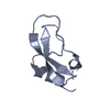
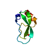
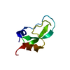
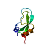
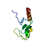
| 登録情報 | データベース: PDB / ID: 1g6x | ||||||
|---|---|---|---|---|---|---|---|
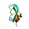
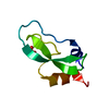
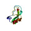
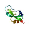
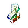
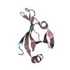
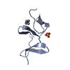
| タイトル | ULTRA HIGH RESOLUTION STRUCTURE OF BOVINE PANCREATIC TRYPSIN INHIBITOR (BPTI) MUTANT WITH ALTERED BINDING LOOP SEQUENCE | ||||||
 要素 要素 | PANCREATIC TRYPSIN INHIBITOR | ||||||
 キーワード キーワード | HYDROLASE INHIBITOR / SERINE PROTEASE INHIBITOR | ||||||
| 機能・相同性 |  機能・相同性情報 機能・相同性情報sulfate binding / negative regulation of platelet aggregation / zymogen binding / potassium channel inhibitor activity / molecular function inhibitor activity / negative regulation of thrombin-activated receptor signaling pathway / serine protease inhibitor complex / serine-type endopeptidase inhibitor activity / protease binding / calcium ion binding / : 類似検索 - 分子機能 | ||||||
| 生物種 |  | ||||||
| 手法 |  X線回折 / シンクロトロン / EXISTING MODEL / 解像度: 0.86 Å X線回折 / シンクロトロン / EXISTING MODEL / 解像度: 0.86 Å | ||||||
 データ登録者 データ登録者 | Addlagatta, A. / Czapinska, H. / Krzywda, S. / Otlewski, J. / Jaskolski, M. | ||||||
 引用 引用 | ジャーナル: Acta Crystallogr.,Sect.D / 年: 2001 タイトル: Ultrahigh-resolution structure of a BPTI mutant. 著者: Addlagatta, A. / Krzywda, S. / Czapinska, H. / Otlewski, J. / Jaskolski, M. #1: ジャーナル: J.Mol.Biol. / 年: 2000タイトル: High-Resolution Structure of Bovine Pancreatic Trypsin Inhibitor with Altered Binding Loop Sequenc 著者: Czapinska, H. / Otlewski, J. / Krzywda, S. / Sheldrick, G.M. / Jaskolski, M. #2: ジャーナル: Acta Crystallogr.,Sect.D / 年: 1996タイトル: Structure of Bovine Pancreatic Trypsin Inhibitor at 125 K: Definition of Carboxyl-Terminal Residues Gly57 and Ala58 著者: Parkin, S. / Rupp, B. / Hope, H. #3: ジャーナル: J.Mol.Biol. / 年: 1992タイトル: Determination of a High Quality Nuclear Magnetic Resonance Solution Structure of the Bovine Pancreatic Trypsin Inhibitor and Comparison with Three Crystal Structures 著者: Berndt, K. / Guentert, P. / Orbons, L.P. / Wuethrich, K. #4: ジャーナル: J.Mol.Biol. / 年: 1984タイトル: Structure of Bovine Pancreatic Trypsin Inhibitor . Results of Joint Neutron and X-Ray Refinement of Crystal Form II 著者: Wlodawer, A. / Walter, J. / Huber, R. / Sjolin, L. #5: ジャーナル: Acta Crystallogr.,Sect.B / 年: 1975タイトル: Crystallographic Refinement of the Structure of Bovine Pancreatic Trypsin Inhibitor at 1.5 A Resolution 著者: Deisenhofer, J. / Steigemann, W. | ||||||
| 履歴 |
|
- 構造の表示
構造の表示
| 構造ビューア | 分子: MolmilJmol/JSmol |
|---|
- ダウンロードとリンク
-ダウンロード
| PDBx/mmCIF形式 | 1g6x.cif.gz | 50.6 KB | 表示 | PDBx/mmCIF形式 |
|---|---|---|---|---|
| PDB形式 | pdb1g6x.ent.gz | 36.6 KB | 表示 | PDB形式 |
| PDBx/mmJSON形式 | 1g6x.json.gz | ツリー表示 | PDBx/mmJSON形式 | |
| その他 |  その他のダウンロード その他のダウンロード |
-検証レポート
| アーカイブディレクトリ | https://data.pdbj.org/pub/pdb/validation_reports/g6/1g6xftp://data.pdbj.org/pub/pdb/validation_reports/g6/1g6x | HTTPS FTP |
|---|
-関連構造データ
| 関連構造データ |  1qlqS S: 精密化の開始モデル |
|---|---|
| 類似構造データ |


-リンク
 PDBj
PDBj


- 集合体
集合体
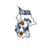
| 登録構造単位 | 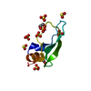
| |||||||||||||||||||||
|---|---|---|---|---|---|---|---|---|---|---|---|---|---|---|---|---|---|---|---|---|---|---|
| 1 |
| |||||||||||||||||||||
| 2 | 
| |||||||||||||||||||||
| 単位格子 |
| |||||||||||||||||||||
| Components on special symmetry positions |
|
-要素
| #1: タンパク質 | 分子量: 6481.481 Da / 分子数: 1 / 変異: YES / 由来タイプ: 組換発現 / 由来: (組換発現)  | ||||||
|---|---|---|---|---|---|---|---|
| #2: 化合物 | ChemComp-SO4 /   分子量: 96.063 Da / 分子数: 8 / 由来タイプ: 合成 / 式: SO4 分子量: 96.063 Da / 分子数: 8 / 由来タイプ: 合成 / 式: SO4#3: 化合物 |   分子量: 62.068 Da / 分子数: 2 / 由来タイプ: 合成 / 式: C2H6O2 分子量: 62.068 Da / 分子数: 2 / 由来タイプ: 合成 / 式: C2H6O2#4: 水 | ChemComp-HOH / |  分子量: 18.015 Da / 分子数: 170 / 由来タイプ: 天然 / 式: H2O 分子量: 18.015 Da / 分子数: 170 / 由来タイプ: 天然 / 式: H2OHas protein modification | Y | |
-実験情報
-実験
| 実験 | 手法: X線回折 / 使用した結晶の数: 1 |
|---|
- 試料調製
試料調製
| 結晶 | マシュー密度: 2.28 Å3/Da / 溶媒含有率: 46 % | |||||||||||||||||||||||||
|---|---|---|---|---|---|---|---|---|---|---|---|---|---|---|---|---|---|---|---|---|---|---|---|---|---|---|
| 結晶化 | 温度: 292 K / 手法: 蒸気拡散法, ハンギングドロップ法 / pH: 7.5 詳細: 2% PEG 400, 2 M AMMONIUM SULFATE, 0.1 M NA HEPES. A PROTEIN SAMPLE, LYOPHILIZED AFTER HPLC PURIFICATION FROM TFA/ACETONITRILE MIXTURE, WAS DISSOLVED IN WATER TO A CONCENTRATION OF 9 MG/2 UL ...詳細: 2% PEG 400, 2 M AMMONIUM SULFATE, 0.1 M NA HEPES. A PROTEIN SAMPLE, LYOPHILIZED AFTER HPLC PURIFICATION FROM TFA/ACETONITRILE MIXTURE, WAS DISSOLVED IN WATER TO A CONCENTRATION OF 9 MG/2 UL DROPS OF THE PROTEIN SOLUTION WERE MIXED WITH 2 UL OF RESERVOIR SOLUTION CONTAINING 2% PEG 400, 2 M AMMONIUM SULFATE AND 0.1 M NA HEPES, PH 7.5. THE HANGING DROPLETS WERE EQUILIBRATED AT 19 DEG C THROUGH THE GAS PHASE WITH THE RESERVOIR. PRISMATIC CRYSTALS MEASURING UP TO 0.4 MM GREW WITHIN 12 HOURS. FOR LOW-TEMPERATURE DATA COLLECTION (100 K), THE CRYSTAL WAS CRYOPROTECTED IN THE RESERVOIR SOLUTION SUPPLEMENTED BY 30 % ETHYLENE GLYCOL., VAPOR DIFFUSION, HANGING DROP, temperature 292K | |||||||||||||||||||||||||
| 結晶化 | *PLUS 温度: 19 ℃ / pH: 7 / 詳細: Czapinska, H., (2000) J.Mol.Biol., 295, 1237. | |||||||||||||||||||||||||
| 溶液の組成 | *PLUS
|
-データ収集
| 回折 | 平均測定温度: 100 K |
|---|---|
| 放射光源 | 由来: シンクロトロン / サイト: EMBL/DESY, HAMBURG  / ビームライン: X11 / 波長: 0.909 / ビームライン: X11 / 波長: 0.909 |
| 検出器 | タイプ: MARRESEARCH / 検出器: IMAGE PLATE / 日付: 1999年7月7日 |
| 放射 | モノクロメーター: SI / プロトコル: SINGLE WAVELENGTH / 単色(M)・ラウエ(L): M / 散乱光タイプ: x-ray |
| 放射波長 | 波長: 0.909 Å / 相対比: 1 |
| 反射 | 解像度: 0.86→20 Å / Num. obs: 47018 / % possible obs: 94.9 % / Observed criterion σ(I): -3 / 冗長度: 17.3 % / Rmerge(I) obs: 0.036 / Net I/σ(I): 54.1 |
| 反射 シェル | 解像度: 0.86→0.9 Å / 冗長度: 6 % / Rmerge(I) obs: 0.488 / Mean I/σ(I) obs: 3.61 / % possible all: 88.6 |
| 反射 | *PLUS Num. measured all: 814213 |
| 反射 シェル | *PLUS % possible obs: 88.6 % |
- 解析
解析
| ソフトウェア |
| |||||||||||||||||||||||||||||||||
|---|---|---|---|---|---|---|---|---|---|---|---|---|---|---|---|---|---|---|---|---|---|---|---|---|---|---|---|---|---|---|---|---|---|---|
| 精密化 | 構造決定の手法: EXISTING MODEL 開始モデル: 1QLQ 解像度: 0.86→10 Å / Num. parameters: 6499 / Num. restraintsaints: 7361 / 交差検証法: FREE R StereochEM target val spec case: ETHYLENE GLYCOL (EDO) AND SULFATE (SO4) GEOMETRY BASED ON DATA FROM CSD 立体化学のターゲット値: ENGH AND HUBER 詳細: ANISOTROPIC REFINEMENT WITHOUT STEREOCHEMICAL RESTRAINTS ON ORDERED MAIN CHAIN. THE COMPLETE C-TERMINUS IS VISIBLE. IT FORMS A SALT-BRIDGE WITH THE N-TERMINUS. ARG 39 IS DISORDERED IN TWO ...詳細: ANISOTROPIC REFINEMENT WITHOUT STEREOCHEMICAL RESTRAINTS ON ORDERED MAIN CHAIN. THE COMPLETE C-TERMINUS IS VISIBLE. IT FORMS A SALT-BRIDGE WITH THE N-TERMINUS. ARG 39 IS DISORDERED IN TWO CONFORMATIONS. IN ADDITION, IT IS ADJACENT TO CYS 38 OF THE DISORDERED 14-38 DISULFIDE AND IS PART OF A DISORDERED ARGININE CAGE. ALA 58 IS THE C-TERMINAL RESIDUE. IT IMMEDIATELY FOLLOWS THE DISORDERED GLY 56 - GLY 57 DOUBLET. THE CYS14-CYS38 DISULFIDE BRIDGE IS OBSERVED IN TWO DISTINCT CHIRALITIES (60 % RIGHT-HANDED, 40 % LEFT-HANDED). THE MAIN CHAIN OF THREE RESIDUES AND THE SIDE CHAINS OF 10 RESIDUES ARE MODELED IN TWO CONFORMATIONS. EIGHT SULFATE ANIONS (TWO WITH TWO-FOLD SYMMETRY) ARE PRESENT IN THE ASYMMETRIC UNIT. ONE OF THEM SHARES A SITE WITH THREE WATER MOLECULES. TWO ETHYLENE GLYCOL MOLECULES MODELED PER ONE PROTEIN MOLECULE. REFINEMENT CONCLUDED USING ONE CYCLE OF BLOCKED FULL-MATRIX LEAST-SQUARES ALGORITHM.
| |||||||||||||||||||||||||||||||||
| 溶媒の処理 | 溶媒モデル: MOEWS & KRETSINGER, J.MOL.BIOL.91(1973)201-228 | |||||||||||||||||||||||||||||||||
| Refine analyze | Num. disordered residues: 12 / Occupancy sum hydrogen: 426 / Occupancy sum non hydrogen: 603 | |||||||||||||||||||||||||||||||||
| 精密化ステップ | サイクル: LAST / 解像度: 0.86→10 Å
| |||||||||||||||||||||||||||||||||
| 拘束条件 |
| |||||||||||||||||||||||||||||||||
| ソフトウェア | *PLUS 名称: SHELXL-97 / 分類: refinement | |||||||||||||||||||||||||||||||||
| 精密化 | *PLUS % reflection Rfree: 4 % / Rfactor Rfree: 0.14 / Rfactor Rwork: 0.107 | |||||||||||||||||||||||||||||||||
| 溶媒の処理 | *PLUS | |||||||||||||||||||||||||||||||||
| 原子変位パラメータ | *PLUS | |||||||||||||||||||||||||||||||||
| 拘束条件 | *PLUS タイプ: s_chiral_restr / Dev ideal: 0.113 |
