 Movie
Movie Controller

[English] 日本語
 Yorodumi
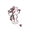
- PDB-1aal: STRUCTURAL EFFECTS INDUCED BY MUTAGENESIS AFFECTED BY CRYSTAL PAC... -
+ Open data

- Basic information
| Entry | Database: PDB / ID: 1aal | ||||||
|---|---|---|---|---|---|---|---|
| Title | STRUCTURAL EFFECTS INDUCED BY MUTAGENESIS AFFECTED BY CRYSTAL PACKING FACTORS: THE STRUCTURE OF A 30-51 DISULFIDE MUTANT OF BASIC PANCREATIC TRYPSIN INHIBITOR | ||||||
 Components Components | BOVINE PANCREATIC TRYPSIN INHIBITOR | ||||||
 Keywords Keywords | SERINE PROTEASE INHIBITOR | ||||||
| Function / homology |  Function and homology information Function and homology informationsulfate binding / negative regulation of platelet aggregation / zymogen binding / potassium channel inhibitor activity / molecular function inhibitor activity / negative regulation of thrombin-activated receptor signaling pathway / serine protease inhibitor complex / serine-type endopeptidase inhibitor activity / protease binding / calcium ion binding / : Similarity search - Function | ||||||
| Biological species |  | ||||||
| Method |  X-RAY DIFFRACTION / Resolution: 1.6 Å X-RAY DIFFRACTION / Resolution: 1.6 Å | ||||||
 Authors Authors | Eigenbrot, C. / Randal, M. / Kossiakoff, A.A. | ||||||
 Citation Citation | Journal: Proteins / Year: 1992 Title: Structural effects induced by mutagenesis affected by crystal packing factors: the structure of a 30-51 disulfide mutant of basic pancreatic trypsin inhibitor. Authors: Eigenbrot, C. / Randal, M. / Kossiakoff, A.A. #1: Journal: Protein Eng. / Year: 1990Title: Structural Effects Induced by Removal of a Disulfide Bridge: The X-Ray Structure of the C30A(Slash)C51A Mutant of Basic Pancreatic Trypsin Inhibitor at 1.6 Angstroms Authors: Eigenbrot, C. / Randal, M. / Kossiakoff, A.A. | ||||||
| History |
|
- Structure visualization
| Structure viewer | Molecule: MolmilJmol/JSmol |
|---|
- Downloads & links
-Download
| PDBx/mmCIF format | 1aal.cif.gz | 38.8 KB | Display | PDBx/mmCIF format |
|---|---|---|---|---|
| PDB format | pdb1aal.ent.gz | 27 KB | Display | PDB format |
| PDBx/mmJSON format | 1aal.json.gz | Tree view | PDBx/mmJSON format | |
| Others |  Other downloads Other downloads |
-Validation report
| Arichive directory | https://data.pdbj.org/pub/pdb/validation_reports/aa/1aalftp://data.pdbj.org/pub/pdb/validation_reports/aa/1aal | HTTPS FTP |
|---|
-Related structure data
| Similar structure data |
|---|


-Links
 PDBj
PDBj


- Assembly
| Deposited unit | 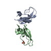
| ||||||||
|---|---|---|---|---|---|---|---|---|---|
| 1 | 
| ||||||||
| 2 | 
| ||||||||
| Unit cell |
| ||||||||
| Atom site foot note | 1: THE FOLLOWING RESIDUES MODELLED WITH 2 SIDE CHAIN CONFORMATIONS: GLU A 7, ARG A 17, ARG A 39, GLU A 49, ASP B 3, ARG B 17, ARG B 39, ARG B 42, GLU B 49. | ||||||||
| Noncrystallographic symmetry (NCS) | NCS oper: (Code: given Matrix: (0.785512, -0.594349, -0.172394), Vector: Details | THE TRANSFORMATION PRESENTED ON *MTRIX* RECORDS BELOW WILL GENERATE APPROXIMATE COORDINATES FOR CHAIN B WHEN APPLIED TO CHAIN A. | |
-Components
| #1: Protein | Mass: 6491.490 Da / Num. of mol.: 2 Source method: isolated from a genetically manipulated source Source: (gene. exp.) #2: Chemical | ChemComp-PO4 / |   Mass: 94.971 Da / Num. of mol.: 1 / Source method: obtained synthetically / Formula: PO4 Mass: 94.971 Da / Num. of mol.: 1 / Source method: obtained synthetically / Formula: PO4#3: Water | ChemComp-HOH / |  Mass: 18.015 Da / Num. of mol.: 126 / Source method: isolated from a natural source / Formula: H2O Mass: 18.015 Da / Num. of mol.: 126 / Source method: isolated from a natural source / Formula: H2OCompound details | THERE IS A UNIQUE SALT-BRIDGE BETWEEN THE N AND C TERMINALS OF MOLECULES WITH RESIDUE NUMBERS 1 - ...THERE IS A UNIQUE SALT-BRIDGE BETWEEN THE N AND C TERMINALS OF MOLECULES WITH RESIDUE NUMBERS 1 - 58, WHICH HAS BEEN SEEN IN SOLUTION (NMR) BUT NEVER CRYSTALLOG | Has protein modification | Y | |
|---|
-Experimental details
-Experiment
| Experiment | Method: X-RAY DIFFRACTION |
|---|
- Sample preparation
| Crystal | Density Matthews: 2.35 Å3/Da / Density % sol: 47.57 % |
|---|---|
| Crystal grow | *PLUS pH: 10 / Method: vapor diffusion, hanging drop |
| Components of the solutions | *PLUS Conc.: 0.7 M / Chemical formula: NaKPO4 |
-Data collection
| Radiation | Scattering type: x-ray |
|---|---|
| Radiation wavelength | Relative weight: 1 |
| Reflection | *PLUS Highest resolution: 1.6 Å / Lowest resolution: 8 Å / Num. obs: 16638 / Rmerge(I) obs: 0.131 |
- Processing
| Software | Name: PROLSQ / Classification: refinement | ||||||||||||||||||||||||||||||||||||||||||||||||||||||||||||||||||||||||||||||||||||
|---|---|---|---|---|---|---|---|---|---|---|---|---|---|---|---|---|---|---|---|---|---|---|---|---|---|---|---|---|---|---|---|---|---|---|---|---|---|---|---|---|---|---|---|---|---|---|---|---|---|---|---|---|---|---|---|---|---|---|---|---|---|---|---|---|---|---|---|---|---|---|---|---|---|---|---|---|---|---|---|---|---|---|---|---|---|
| Refinement | Resolution: 1.6→8 Å / σ(I): 1 /
| ||||||||||||||||||||||||||||||||||||||||||||||||||||||||||||||||||||||||||||||||||||
| Refinement step | Cycle: LAST / Resolution: 1.6→8 Å
| ||||||||||||||||||||||||||||||||||||||||||||||||||||||||||||||||||||||||||||||||||||
| Refine LS restraints |
| ||||||||||||||||||||||||||||||||||||||||||||||||||||||||||||||||||||||||||||||||||||
| Refinement | *PLUS Highest resolution: 1.6 Å / Lowest resolution: 8 Å / Num. reflection obs: 15473 / σ(I): 1 / Rfactor obs: 0.179 | ||||||||||||||||||||||||||||||||||||||||||||||||||||||||||||||||||||||||||||||||||||
| Solvent computation | *PLUS | ||||||||||||||||||||||||||||||||||||||||||||||||||||||||||||||||||||||||||||||||||||
| Displacement parameters | *PLUS |
