 Movie
Movie Controller

[English] 日本語
 Yorodumi
- PDB-2zvs: Crystal structure of the 2[4FE-4S] ferredoxin from escherichia coli -
+ Open data

- Basic information
| Entry | Database: PDB / ID: 2zvs | ||||||
|---|---|---|---|---|---|---|---|
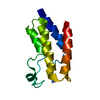
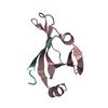
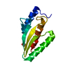
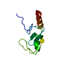
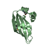
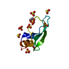
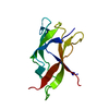
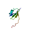
| Title | Crystal structure of the 2[4FE-4S] ferredoxin from escherichia coli | ||||||
 Components Components | Uncharacterized ferredoxin-like protein yfhL | ||||||
 Keywords Keywords | ELECTRON TRANSPORT / FERREDOXIN / [4FE-4S] CLUSTERS / IRON-SULFUR CLUSTERS / ESCHERICHIA COLI / reduction potential / Iron binding protein / Iron / Metal-binding | ||||||
| Function / homology |  Function and homology information Function and homology informationtRNA wobble base modification / 4 iron, 4 sulfur cluster binding / metal ion binding / cytoplasm Similarity search - Function | ||||||
| Biological species |  | ||||||
| Method |  X-RAY DIFFRACTION / SYNCHROTRON / MOLECULAR REPLACEMENT / Resolution: 1.65 Å X-RAY DIFFRACTION / SYNCHROTRON / MOLECULAR REPLACEMENT / Resolution: 1.65 Å | ||||||
 Authors Authors | Giastas, P. / Mavridis, M.I. | ||||||
 Citation Citation | Journal: J.Biol.Inorg.Chem. / Year: 2009 Title: Insight into the protein and solvent contributions to the reduction potentials of [4Fe-4S]2+/+ clusters: crystal structures of the Allochromatium vinosum ferredoxin variants C57A and V13G and ...Title: Insight into the protein and solvent contributions to the reduction potentials of [4Fe-4S]2+/+ clusters: crystal structures of the Allochromatium vinosum ferredoxin variants C57A and V13G and the homologous Escherichia coli ferredoxin Authors: Saridakis, E. / Giastas, P. / Efthymiou, G. / Thoma, V. / Moulis, J.M. / Kyritsis, P. / Mavridis, I.M. | ||||||
| History |
|
- Structure visualization
| Structure viewer | Molecule: MolmilJmol/JSmol |
|---|
- Downloads & links
-Download
| PDBx/mmCIF format | 2zvs.cif.gz | 70.4 KB | Display | PDBx/mmCIF format |
|---|---|---|---|---|
| PDB format | pdb2zvs.ent.gz | 50.6 KB | Display | PDB format |
| PDBx/mmJSON format | 2zvs.json.gz | Tree view | PDBx/mmJSON format | |
| Others |  Other downloads Other downloads |
-Validation report
| Arichive directory | https://data.pdbj.org/pub/pdb/validation_reports/zv/2zvsftp://data.pdbj.org/pub/pdb/validation_reports/zv/2zvs | HTTPS FTP |
|---|
-Related structure data
| Related structure data | 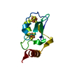 3eunC 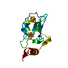 3exyC  1bluS S: Starting model for refinement C: citing same article ( |
|---|---|
| Similar structure data |


-Links
 PDBj
PDBj


- Assembly
| Deposited unit | 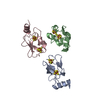
| |||||||||
|---|---|---|---|---|---|---|---|---|---|---|
| 1 | 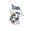
| |||||||||
| 2 | 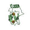
| |||||||||
| 3 | 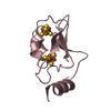
| |||||||||
| Unit cell |
| |||||||||
| Components on special symmetry positions |
| |||||||||
| Details | the biological unit is unknown. |
-Components
| #1: Protein | Mass: 9672.118 Da / Num. of mol.: 3 Source method: isolated from a genetically manipulated source Source: (gene. exp.) #2: Chemical | ChemComp-SF4 /   Mass: 351.640 Da / Num. of mol.: 6 / Source method: obtained synthetically / Formula: Fe4S4 Mass: 351.640 Da / Num. of mol.: 6 / Source method: obtained synthetically / Formula: Fe4S4#3: Water | ChemComp-HOH / |  Mass: 18.015 Da / Num. of mol.: 179 / Source method: isolated from a natural source / Formula: H2O Mass: 18.015 Da / Num. of mol.: 179 / Source method: isolated from a natural source / Formula: H2O |
|---|
-Experimental details
-Experiment
| Experiment | Method: X-RAY DIFFRACTION / Number of used crystals: 1 |
|---|
- Sample preparation
| Crystal | Density Matthews: 2.83 Å3/Da / Density % sol: 56.52 % / Description: The file contains Friedel pair. |
|---|---|
| Crystal grow | Temperature: 277 K / Method: vapor diffusion, hanging drop / pH: 9 Details: 0.5M CACL2, 0.1M TRIS-HCL, 20% PEG 4000, pH 9.0, VAPOR DIFFUSION, HANGING DROP, temperature 277K |
-Data collection
| Diffraction | Mean temperature: 100 K |
|---|---|
| Diffraction source | Source: SYNCHROTRON / Site: EMBL/DESY, HAMBURG  / Beamline: X13 / Wavelength: 0.81 Å / Beamline: X13 / Wavelength: 0.81 Å |
| Detector | Type: MARRESEARCH / Detector: CCD / Date: Jul 25, 2005 |
| Radiation | Protocol: SINGLE WAVELENGTH / Monochromatic (M) / Laue (L): M / Scattering type: x-ray |
| Radiation wavelength | Wavelength: 0.81 Å / Relative weight: 1 |
| Reflection | Resolution: 1.65→40 Å / Num. obs: 71543 / Observed criterion σ(F): 4 / Redundancy: 5.8 % / Biso Wilson estimate: 17 Å2 / Rmerge(I) obs: 0.066 |
| Reflection shell | Resolution: 1.65→1.68 Å / Mean I/σ(I) obs: 2 / % possible all: 100 |
- Processing
| Software |
| |||||||||||||||||||||||||||||||||
|---|---|---|---|---|---|---|---|---|---|---|---|---|---|---|---|---|---|---|---|---|---|---|---|---|---|---|---|---|---|---|---|---|---|---|
| Refinement | Method to determine structure: MOLECULAR REPLACEMENT Starting model: 1BLU Resolution: 1.65→30 Å / Num. parameters: 8484 / Num. restraintsaints: 8348 / Isotropic thermal model: ISOTROPIC / Cross valid method: FREE R / σ(F): 4 / Stereochemistry target values: Engh & Huber Details: The file contains Friedel pair. Used weighted CONJUGATED GRADIENT MATRIX least squares procedure.
| |||||||||||||||||||||||||||||||||
| Refine analyze | Num. disordered residues: 0 / Occupancy sum hydrogen: 0 / Occupancy sum non hydrogen: 2042 | |||||||||||||||||||||||||||||||||
| Refinement step | Cycle: LAST / Resolution: 1.65→30 Å
| |||||||||||||||||||||||||||||||||
| Refine LS restraints |
|
