[English] 日本語
Yorodumi
- PDB-1zv6: NMR structure of the human dematin headpiece S74E mutant -

+
Open data


ID or keywords:

Loading...

-
Basic information

Entry
Database: PDB / ID: 1zv6
TitleNMR structure of the human dematin headpiece S74E mutant
ComponentsEPB49 protein
KeywordsPROTEIN BINDING / dematin headpiece / actin binding domain / phosphorylation
Function / homology
Function and homology information


negative regulation of protein targeting to membrane / spectrin-associated cytoskeleton / negative regulation of peptidyl-tyrosine phosphorylation / negative regulation of substrate adhesion-dependent cell spreading / Miscellaneous transport and binding events / platelet dense tubular network membrane / negative regulation of focal adhesion assembly / cell projection membrane / regulation of filopodium assembly / actin filament capping ...negative regulation of protein targeting to membrane / spectrin-associated cytoskeleton / negative regulation of peptidyl-tyrosine phosphorylation / negative regulation of substrate adhesion-dependent cell spreading / Miscellaneous transport and binding events / platelet dense tubular network membrane / negative regulation of focal adhesion assembly / cell projection membrane / regulation of filopodium assembly / actin filament capping / positive regulation of fibroblast migration / regulation of lamellipodium assembly / negative regulation of cell-substrate adhesion / lamellipodium assembly / spectrin binding / positive regulation of wound healing / cortical cytoskeleton / negative regulation of peptidyl-threonine phosphorylation / negative regulation of peptidyl-serine phosphorylation / actin filament bundle assembly / : / positive regulation of blood coagulation / endomembrane system / erythrocyte development / cellular response to cAMP / cytoskeleton organization / cellular response to calcium ion / actin filament / regulation of actin cytoskeleton organization / : / actin filament binding / actin cytoskeleton / actin binding / regulation of cell shape / cytoplasmic vesicle / actin cytoskeleton organization / protein-containing complex assembly / postsynaptic density / signaling receptor binding / perinuclear region of cytoplasm / plasma membrane / cytosol
Similarity search - Function
Putative adherens-junction anchoring domain / Putative adherens-junction anchoring region of AbLIM / Villin Headpiece Domain; Chain A / Villin headpiece domain / Villin headpiece / Villin headpiece domain superfamily / Villin headpiece domain / Headpiece (HP) domain profile. / Villin headpiece domain / Orthogonal Bundle / Mainly Alpha
Similarity search - Domain/homology
Biological speciesHomo sapiens (human)
MethodSOLUTION NMR / DISTANCE GEOMETRY, SIMULATED ANNEALING
Model type detailsminimized average
AuthorsJiang, Z.G. / McKnight, C.J.
CitationJournal: Structure / Year: 2006
Title: A phosphorylation-induced conformation change in dematin headpiece.
Authors: Jiang, Z.G. / McKnight, C.J.
History
DepositionJun 1, 2005Deposition site: RCSB / Processing site: RCSB
Revision 1.0Mar 21, 2006Provider: repository / Type: Initial release
Revision 1.1Apr 30, 2008Group: Version format compliance
Revision 1.2Jul 13, 2011Group: Version format compliance
Revision 1.3Oct 20, 2021Group: Data collection / Database references / Derived calculations
Category: database_2 / pdbx_nmr_software ...database_2 / pdbx_nmr_software / pdbx_struct_assembly / pdbx_struct_oper_list / struct_ref_seq_dif
Item: _database_2.pdbx_DOI / _database_2.pdbx_database_accession ..._database_2.pdbx_DOI / _database_2.pdbx_database_accession / _pdbx_nmr_software.name / _struct_ref_seq_dif.details
Revision 1.4May 22, 2024Group: Data collection / Category: chem_comp_atom / chem_comp_bond
Remark 999
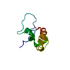
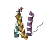
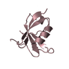
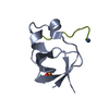
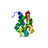
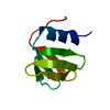
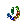
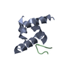
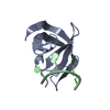
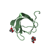
SEQUENCE THE DHPS74E CONSTRUCT SPANS RESIDUES 316 TO 383 OF THE 48 KDA FORM OF HUMAN DEMATIN, WITH ...SEQUENCE THE DHPS74E CONSTRUCT SPANS RESIDUES 316 TO 383 OF THE 48 KDA FORM OF HUMAN DEMATIN, WITH SER74 REPLACED BY GLU. TO FACILITATE THE COMPARISON WITH OTHER HEADPIECE DOMAINS, WE USE THE NUMBERING SCHEME FROM VARDAR ET AL., 1999, J.MOLEC.BIOL., 294, 1299-1310, SUCH THAT THE N-TERMINAL PROLINE OF DHP IS RESIDUE 9, AND C-TERMINAL PHENYLALANINE IS RESIDUE 76. THE FINAL RESIDUE (F76) IS THE NATURAL C-TERMINAL RESIDUE OF DEMATIN.

-
Structure visualization

Structure viewerMolecule:
MolmilJmol/JSmol

Downloads & links

-
Assembly

Deposited unit
A: EPB49 protein


Theoretical massNumber of molelcules
Total (without water)7,9781
Polymers7,9781
Non-polymers00
Water00
1


  • Idetical with deposited unit
  • defined by author
TypeNameSymmetry operationNumber
identity operation1_5551
NMR ensembles
DataCriteria
Number of conformers (submitted / calculated)21 / 100structures with the lowest energy
RepresentativeModel #1minimized average structure

-
Components

#1: Protein EPB49 protein


Mass: 7978.362 Da / Num. of mol.: 1 / Fragment: residues 316-383
Source method: isolated from a genetically manipulated source
Source: (gene. exp.) Homo sapiens (human) / Plasmid: PD48 / Species (production host): Escherichia coli / Production host: Escherichia coli BL21(DE3) (bacteria) / Strain (production host): BL21(DE3) / References: GenBank: 31127246, UniProt: Q08495*PLUS

-
Experimental details

-
Experiment

ExperimentMethod: SOLUTION NMR
NMR experiment
Conditions-IDExperiment-IDSolution-IDType
1113D 13C-separated NOESY
121HNCA-J
131HN(CO)CA
1412D NOESY
1513D 15N-separated NOESY
161HNHA
171HNHB
1812D NOESY
1912D TOCSY
NMR detailsText: THE STRUCTURE WAS DETERMINED USING TRIPLE-RESONANCE NMR SPECTROSCOPY. Model 1 represents minimized average structure.

-
Sample preparation

DetailsContents: 1 MM DHPs74e U-15N,13C U-15N; 1 MM DHPs74e 10% 13C, U- 15N, 90% H2O, 10% D2O
Solvent system: 90% H2O/10% D2O
Sample conditionsIonic strength: 10 mM PHOSPHATE / pH: 6.0 / Pressure: AMBIENT / Temperature: 293 K

-
NMR measurement

RadiationProtocol: SINGLE WAVELENGTH / Monochromatic (M) / Laue (L): M
Radiation wavelengthRelative weight: 1
NMR spectrometerType: Bruker DMX / Manufacturer: Bruker / Model: DMX / Field strength: 500 MHz

-
Processing

NMR software
NameVersionDeveloperClassification
MOLMOL2K.2KORADIstructure solution
CNS1Brungerrefinement
NMRPipe2004 versionDELAGLIOprocessing
XwinNMR3.1collection
NMRView5.0.4.Johnsondata analysis
RefinementMethod: DISTANCE GEOMETRY, SIMULATED ANNEALING / Software ordinal: 1
Details: THE STRUCTURES ARE BASED ON A TOTAL OF 1449 RESTRAINTS: 1289 ARE NOE-DERIVED DISTANCE CONSTRAINTS, 142 ARE DIHEDRAL ANGLE RESTRAINTS, AND 18 ARE DISTANCE RESTRAINTS FROM HYDROGEN BONDS.
NMR representativeSelection criteria: minimized average structure
NMR ensembleConformer selection criteria: structures with the lowest energy
Conformers calculated total number: 100 / Conformers submitted total number: 21

+
About Yorodumi

-
News

-
Feb 9, 2022. New format data for meta-information of EMDB entries

New format data for meta-information of EMDB entries

  • Version 3 of the EMDB header file is now the official format.
  • The previous official version 1.9 will be removed from the archive.

Related info.:EMDB header

External links:wwPDB to switch to version 3 of the EMDB data model

-
Aug 12, 2020. Covid-19 info

Covid-19 info

URL: https://pdbjlvh1.pdbj.org/emnavi/covid19.php

New page: Covid-19 featured information page in EM Navigator.

Related info.:Covid-19 info / Mar 5, 2020. Novel coronavirus structure data

+
Mar 5, 2020. Novel coronavirus structure data

Novel coronavirus structure data

Related info.:Yorodumi Speices / Aug 12, 2020. Covid-19 info

External links:COVID-19 featured content - PDBj / Molecule of the Month (242):Coronavirus Proteases

+
Jan 31, 2019. EMDB accession codes are about to change! (news from PDBe EMDB page)

EMDB accession codes are about to change! (news from PDBe EMDB page)

  • The allocation of 4 digits for EMDB accession codes will soon come to an end. Whilst these codes will remain in use, new EMDB accession codes will include an additional digit and will expand incrementally as the available range of codes is exhausted. The current 4-digit format prefixed with “EMD-” (i.e. EMD-XXXX) will advance to a 5-digit format (i.e. EMD-XXXXX), and so on. It is currently estimated that the 4-digit codes will be depleted around Spring 2019, at which point the 5-digit format will come into force.
  • The EM Navigator/Yorodumi systems omit the EMD- prefix.

Related info.:Q: What is EMD? / ID/Accession-code notation in Yorodumi/EM Navigator

External links:EMDB Accession Codes are Changing Soon! / Contact to PDBj

+
Jul 12, 2017. Major update of PDB

Major update of PDB

  • wwPDB released updated PDB data conforming to the new PDBx/mmCIF dictionary.
  • This is a major update changing the version number from 4 to 5, and with Remediation, in which all the entries are updated.
  • In this update, many items about electron microscopy experimental information are reorganized (e.g. em_software).
  • Now, EM Navigator and Yorodumi are based on the updated data.

External links:wwPDB Remediation / Enriched Model Files Conforming to OneDep Data Standards Now Available in the PDB FTP Archive

-
Yorodumi

Thousand views of thousand structures

  • Yorodumi is a browser for structure data from EMDB, PDB, SASBDB, etc.
  • This page is also the successor to EM Navigator detail page, and also detail information page/front-end page for Omokage search.
  • The word "yorodu" (or yorozu) is an old Japanese word meaning "ten thousand". "mi" (miru) is to see.

Related info.:EMDB / PDB / SASBDB / Comparison of 3 databanks / Yorodumi Search / Aug 31, 2016. New EM Navigator & Yorodumi / Yorodumi Papers / Jmol/JSmol / Function and homology information / Changes in new EM Navigator and Yorodumi

Read more