 Movie
Movie Controller


+ Open data

- Basic information
| Entry | Database: PDB / ID: 1zsz | ||||||
|---|---|---|---|---|---|---|---|
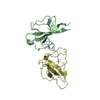
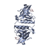
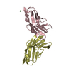
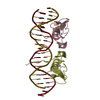
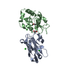
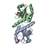
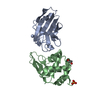
| Title | Crystal structure of a computationally designed SspB heterodimer | ||||||
 Components Components | (Stringent starvation protein B homolog) x 3 | ||||||
 Keywords Keywords | DE NOVO PROTEIN / protein design / AAA / adaptor / specificity | ||||||
| Function / homology |  Function and homology information Function and homology information | ||||||
| Biological species |  Haemophilus influenzae (bacteria) Haemophilus influenzae (bacteria) | ||||||
| Method |  X-RAY DIFFRACTION / SYNCHROTRON / MOLECULAR REPLACEMENT / Resolution: 2 Å X-RAY DIFFRACTION / SYNCHROTRON / MOLECULAR REPLACEMENT / Resolution: 2 Å | ||||||
 Authors Authors | Bolon, D.N. / Grant, R.A. / Baker, T.A. / Sauer, R.T. | ||||||
 Citation Citation | Journal: Proc.Natl.Acad.Sci.Usa / Year: 2005 Title: Specificity versus stability in computational protein design. Authors: Bolon, D.N. / Grant, R.A. / Baker, T.A. / Sauer, R.T. | ||||||
| History |
|
- Structure visualization
| Structure viewer | Molecule: MolmilJmol/JSmol |
|---|
- Downloads & links
-Download
| PDBx/mmCIF format | 1zsz.cif.gz | 78.4 KB | Display | PDBx/mmCIF format |
|---|---|---|---|---|
| PDB format | pdb1zsz.ent.gz | 58.2 KB | Display | PDB format |
| PDBx/mmJSON format | 1zsz.json.gz | Tree view | PDBx/mmJSON format | |
| Others |  Other downloads Other downloads |
-Validation report
| Arichive directory | https://data.pdbj.org/pub/pdb/validation_reports/zs/1zszftp://data.pdbj.org/pub/pdb/validation_reports/zs/1zsz | HTTPS FTP |
|---|
-Related structure data
| Related structure data | 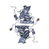 1ou9S S: Starting model for refinement |
|---|---|
| Similar structure data |


-Links
 PDBj
PDBj
- Assembly
| Deposited unit | 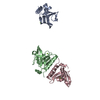
| ||||||||
|---|---|---|---|---|---|---|---|---|---|
| 1 | 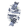
| ||||||||
| 2 | 
| ||||||||
| 3 | 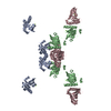
| ||||||||
| Unit cell |
| ||||||||
| Details | chain b and c compose one biological unit, while chain A, forms a dimer across a crystal axis (to a crystallographically related chain A) |
-Components
| #1: Protein | Mass: 12511.090 Da / Num. of mol.: 1 Source method: isolated from a genetically manipulated source Source: (gene. exp.) Haemophilus influenzae (bacteria) / Gene: sspB / Plasmid: pET21, pACYCDuet1 / Production host: | ||
|---|---|---|---|
| #2: Protein | Mass: 12578.135 Da / Num. of mol.: 1 / Mutation: A15S,Y16L,V101A Source method: isolated from a genetically manipulated source Source: (gene. exp.) Haemophilus influenzae (bacteria) / Gene: sspB / Plasmid: pET21 / Production host: | ||
| #3: Protein | Mass: 14769.420 Da / Num. of mol.: 1 / Mutation: L12Y,A15G,Y16F,V101M Source method: isolated from a genetically manipulated source Source: (gene. exp.) Haemophilus influenzae (bacteria) / Gene: sspB / Plasmid: pACYCDuet1 / Production host: | ||
| #4: Chemical |   Mass: 24.305 Da / Num. of mol.: 3 / Source method: obtained synthetically / Formula: Mg Mass: 24.305 Da / Num. of mol.: 3 / Source method: obtained synthetically / Formula: Mg#5: Water | ChemComp-HOH / |  Mass: 18.015 Da / Num. of mol.: 93 / Source method: isolated from a natural source / Formula: H2O Mass: 18.015 Da / Num. of mol.: 93 / Source method: isolated from a natural source / Formula: H2O |
-Experimental details
-Experiment
| Experiment | Method: X-RAY DIFFRACTION / Number of used crystals: 1 |
|---|
- Sample preparation
| Crystal | Density Matthews: 2.68 Å3/Da / Density % sol: 54.05 % |
|---|---|
| Crystal grow | Temperature: 293 K / Method: vapor diffusion, hanging drop / pH: 5.7 Details: PEG 6000, CaCl2, sodium cacodylate, KCl, glycerol, pH 5.7, VAPOR DIFFUSION, HANGING DROP, temperature 293K |
-Data collection
| Diffraction | Mean temperature: 100 K |
|---|---|
| Diffraction source | Source: SYNCHROTRON / Site: APS  / Beamline: 8-BM / Wavelength: 0.97949 Å / Beamline: 8-BM / Wavelength: 0.97949 Å |
| Detector | Type: ADSC QUANTUM 315 / Detector: CCD / Date: Aug 1, 2004 |
| Radiation | Monochromator: SILICON CRYSTAL / Protocol: SINGLE WAVELENGTH / Monochromatic (M) / Laue (L): M / Scattering type: x-ray |
| Radiation wavelength | Wavelength: 0.97949 Å / Relative weight: 1 |
| Reflection | Resolution: 2→30 Å / Num. all: 28612 / Num. obs: 28612 / % possible obs: 100 % / Observed criterion σ(F): 0 / Observed criterion σ(I): 0 / Redundancy: 7.2 % / Biso Wilson estimate: 26.5 Å2 / Rmerge(I) obs: 0.086 |
| Reflection shell | Resolution: 2→2.07 Å / Redundancy: 5.9 % / Rmerge(I) obs: 0.371 / % possible all: 99.8 |
- Processing
| Software |
| |||||||||||||||||||||||||
|---|---|---|---|---|---|---|---|---|---|---|---|---|---|---|---|---|---|---|---|---|---|---|---|---|---|---|
| Refinement | Method to determine structure: MOLECULAR REPLACEMENT Starting model: 1ou9 chain A Resolution: 2→29.15 Å / Rfactor Rfree error: 0.007 / Data cutoff high absF: 432252.67 / Data cutoff high rms absF: 432252.67 / Data cutoff low absF: 0 / Isotropic thermal model: RESTRAINED / Cross valid method: THROUGHOUT / σ(F): 0 / Stereochemistry target values: Engh & Huber Details: Chain A forms a dimer across a crystal axis and is an average of chains B and C. During refinement, residues which are different in chains B and C, were modeled with truncated side chains in ...Details: Chain A forms a dimer across a crystal axis and is an average of chains B and C. During refinement, residues which are different in chains B and C, were modeled with truncated side chains in chain A. The side chains for these residues are present in the chains B and C. The B/C dimer shows the asymmetric packing at the dimer interface that was introduced by computational design and was the focus of this study.
| |||||||||||||||||||||||||
| Solvent computation | Solvent model: FLAT MODEL / Bsol: 41.2607 Å2 / ksol: 0.352752 e/Å3 | |||||||||||||||||||||||||
| Displacement parameters | Biso mean: 41.7 Å2
| |||||||||||||||||||||||||
| Refine analyze |
| |||||||||||||||||||||||||
| Refinement step | Cycle: LAST / Resolution: 2→29.15 Å
| |||||||||||||||||||||||||
| Refine LS restraints |
| |||||||||||||||||||||||||
| LS refinement shell | Resolution: 2→2.13 Å / Rfactor Rfree error: 0.023 / Total num. of bins used: 6
| |||||||||||||||||||||||||
| Xplor file |
|
