 Movie
Movie Controller

[English] 日本語
 Yorodumi
- PDB-1zof: Crystal structure of alkyl hydroperoxide-reductase (AhpC) from He... -
+ Open data

- Basic information
| Entry | Database: PDB / ID: 1zof | ||||||
|---|---|---|---|---|---|---|---|
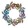
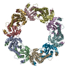
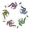
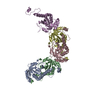
| Title | Crystal structure of alkyl hydroperoxide-reductase (AhpC) from Helicobacter Pylori | ||||||
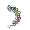
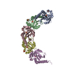
 Components Components | alkyl hydroperoxide-reductase | ||||||
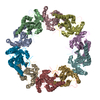
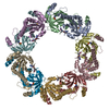
 Keywords Keywords | OXIDOREDUCTASE / decamer / toroide-shaped complex | ||||||
| Function / homology |  Function and homology information Function and homology informationNADH-dependent peroxiredoxin / NADH-dependent peroxiredoxin activity / thioredoxin peroxidase activity / cellular response to stress / cell redox homeostasis / hydrogen peroxide catabolic process / response to oxidative stress / cytosol Similarity search - Function | ||||||
| Biological species |   Helicobacter pylori (bacteria) Helicobacter pylori (bacteria) | ||||||
| Method |  X-RAY DIFFRACTION / SYNCHROTRON / MOLECULAR REPLACEMENT / Resolution: 2.95 Å X-RAY DIFFRACTION / SYNCHROTRON / MOLECULAR REPLACEMENT / Resolution: 2.95 Å | ||||||
 Authors Authors | Papinutto, E. / Windle, H.J. / Cendron, L. / Battistutta, R. / Kelleher, D. / Zanotti, G. | ||||||
 Citation Citation | Journal: Biochim.Biophys.Acta / Year: 2005 Title: Crystal structure of alkyl hydroperoxide-reductase (AhpC) from Helicobacter pylori. Authors: Papinutto, E. / Windle, H.J. / Cendron, L. / Battistutta, R. / Kelleher, D. / Zanotti, G. #1: Journal: J.Mol.Biol. / Year: 2000Title: The structure of reduced tryparedoxin peroxidase reveals a decamer and insight into reactivity of 2Cys-peroxiredoxins Authors: Alphey, M.S. / Bond, C.S. / Tetand, E. / Fairlamb, A.H. / Hunter, W.N. #2: Journal: J.Bacteriol. / Year: 2001Title: Essential thioredoxin-dependent peroxiredoxin system from Helicobacter pylori: genetic and kinetic characterization Authors: Baker, L.M.S. / Raudonikiene, A. / Hoffman, P.S. / Poole, L.B. #3: Journal: TRENDS BIOCHEM.SCI. / Year: 2003Title: Structure, mechanism and regulation of peroxiredoxins Authors: Wood, Z.A. / Schroder, E. / Harris, J.R. / Poole, L.B. | ||||||
| History |
|
- Structure visualization
| Structure viewer | Molecule: MolmilJmol/JSmol |
|---|
- Downloads & links
-Download
| PDBx/mmCIF format | 1zof.cif.gz | 340.6 KB | Display | PDBx/mmCIF format |
|---|---|---|---|---|
| PDB format | pdb1zof.ent.gz | 279.7 KB | Display | PDB format |
| PDBx/mmJSON format | 1zof.json.gz | Tree view | PDBx/mmJSON format | |
| Others |  Other downloads Other downloads |
-Validation report
| Arichive directory | https://data.pdbj.org/pub/pdb/validation_reports/zo/1zofftp://data.pdbj.org/pub/pdb/validation_reports/zo/1zof | HTTPS FTP |
|---|
-Related structure data
| Related structure data |  1e2yS S: Starting model for refinement |
|---|---|
| Similar structure data |

-Links
 PDBj
PDBj- Assembly
| Deposited unit | 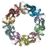
| ||||||||
|---|---|---|---|---|---|---|---|---|---|
| 1 |
| ||||||||
| Unit cell |
| ||||||||
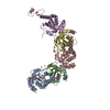
| Details | The biological assembly is a decamer in the asymmetric unit |
-Components
| #1: Protein | Mass: 22276.652 Da / Num. of mol.: 10 / Mutation: V2L Source method: isolated from a genetically manipulated source Source: (gene. exp.) Helicobacter pylori (bacteria) / Gene: tsaA / Plasmid: PBAD-THIO-TOPO / Production host: #2: Water | ChemComp-HOH / |  Mass: 18.015 Da / Num. of mol.: 65 / Source method: isolated from a natural source / Formula: H2O Mass: 18.015 Da / Num. of mol.: 65 / Source method: isolated from a natural source / Formula: H2OHas protein modification | Y | |
|---|
-Experimental details
-Experiment
| Experiment | Method: X-RAY DIFFRACTION / Number of used crystals: 1 |
|---|
- Sample preparation
| Crystal | Density Matthews: 3.05 Å3/Da / Density % sol: 60 % |
|---|---|
| Crystal grow | Temperature: 293 K / Method: vapor diffusion / pH: 4.5 Details: sodium acetate, sodium formate, pH 4.5, VAPOR DIFFUSION, temperature 293K |
-Data collection
| Diffraction | Mean temperature: 100 K |
|---|---|
| Diffraction source | Source: SYNCHROTRON / Site: ESRF  / Beamline: ID29 / Wavelength: 1.27 Å / Beamline: ID29 / Wavelength: 1.27 Å |
| Detector | Type: ADSC QUANTUM 4 / Detector: CCD / Date: Jul 17, 2004 / Details: mirrors |
| Radiation | Monochromator: Si(111) monochromator / Protocol: SINGLE WAVELENGTH / Monochromatic (M) / Laue (L): M / Scattering type: x-ray |
| Radiation wavelength | Wavelength: 1.27 Å / Relative weight: 1 |
| Reflection | Resolution: 2.95→52.8 Å / Num. all: 57514 / Num. obs: 57514 / % possible obs: 99.9 % / Observed criterion σ(F): 0 / Observed criterion σ(I): 0 / Redundancy: 3.1 % / Biso Wilson estimate: 157.9 Å2 / Rmerge(I) obs: 0.091 / Net I/σ(I): 7 |
| Reflection shell | Resolution: 2.95→3.11 Å / Redundancy: 3.1 % / Rmerge(I) obs: 0.39 / Mean I/σ(I) obs: 1.6 / Num. unique all: 8357 / % possible all: 100 |
- Processing
| Software |
| |||||||||||||||||||||||||
|---|---|---|---|---|---|---|---|---|---|---|---|---|---|---|---|---|---|---|---|---|---|---|---|---|---|---|
| Refinement | Method to determine structure: MOLECULAR REPLACEMENT Starting model: PDB ENTRY 1E2Y Resolution: 2.95→52.8 Å / Rfactor Rfree error: 0.004 / Data cutoff high absF: 737506.01 / Data cutoff low absF: 0 / Isotropic thermal model: RESTRAINED / Cross valid method: THROUGHOUT / σ(F): 0 / Stereochemistry target values: Engh & Huber
| |||||||||||||||||||||||||
| Solvent computation | Solvent model: FLAT MODEL / Bsol: 54.6117 Å2 / ksol: 0.369771 e/Å3 | |||||||||||||||||||||||||
| Displacement parameters | Biso mean: 59.6 Å2
| |||||||||||||||||||||||||
| Refine analyze |
| |||||||||||||||||||||||||
| Refinement step | Cycle: LAST / Resolution: 2.95→52.8 Å
| |||||||||||||||||||||||||
| Refine LS restraints |
| |||||||||||||||||||||||||
| Refine LS restraints NCS | NCS model details: CONSTR | |||||||||||||||||||||||||
| LS refinement shell | Resolution: 2.95→3.13 Å / Rfactor Rfree error: 0.014 / Total num. of bins used: 6
| |||||||||||||||||||||||||
| Xplor file |
|
