 Movie
Movie Controller

[English] 日本語
 Yorodumi
- PDB-1zn1: Coordinates of RRF fitted into Cryo-EM map of the 70S post-termin... -
+ Open data

- Basic information
| Entry | Database: PDB / ID: 1zn1 | ||||||
|---|---|---|---|---|---|---|---|
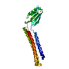
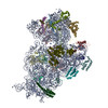
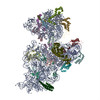
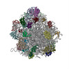
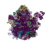
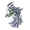
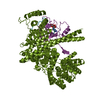
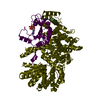
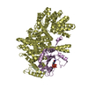
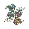
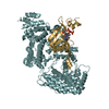
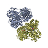
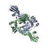
| Title | Coordinates of RRF fitted into Cryo-EM map of the 70S post-termination complex | ||||||
 Components Components |
| ||||||
 Keywords Keywords | BIOSYNTHETIC/structural protein/RNA / ribosome recycling factor / elongation factor G / 70S / post-termination complex / BIOSYNTHETIC-structural protein-RNA COMPLEX | ||||||
| Function / homology |  Function and homology information Function and homology informationcytoplasmic translational termination / ribosomal large subunit binding / misfolded RNA binding / Group I intron splicing / RNA folding / positive regulation of RNA splicing / maintenance of translational fidelity / cytosolic small ribosomal subunit / cytoplasmic translation / tRNA binding ...cytoplasmic translational termination / ribosomal large subunit binding / misfolded RNA binding / Group I intron splicing / RNA folding / positive regulation of RNA splicing / maintenance of translational fidelity / cytosolic small ribosomal subunit / cytoplasmic translation / tRNA binding / rRNA binding / structural constituent of ribosome / ribosome / translation / response to antibiotic / cytoplasm / cytosol Similarity search - Function | ||||||
| Biological species |  | ||||||
| Method | ELECTRON MICROSCOPY / single particle reconstruction / cryo EM / Resolution: 14.1 Å | ||||||
 Authors Authors | Gao, N. / Zavialov, A.V. / Li, W. / Sengupta, J. / Valle, M. / Gursky, R.P. / Ehrenberg, M. / Frank, J. | ||||||
 Citation Citation | Journal: Mol Cell / Year: 2005 Title: Mechanism for the disassembly of the posttermination complex inferred from cryo-EM studies. Authors: Ning Gao / Andrey V Zavialov / Wen Li / Jayati Sengupta / Mikel Valle / Richard P Gursky / Måns Ehrenberg / Joachim Frank /  Abstract: Ribosome recycling, the disassembly of the posttermination complex after each round of protein synthesis, is an essential step in mRNA translation, but its mechanism has remained obscure. In ...Ribosome recycling, the disassembly of the posttermination complex after each round of protein synthesis, is an essential step in mRNA translation, but its mechanism has remained obscure. In eubacteria, recycling is catalyzed by RRF (ribosome recycling factor) and EF-G (elongation factor G). By using cryo-electron microscopy, we have obtained two density maps, one of the RRF bound posttermination complex and one of the 50S subunit bound with both EF-G and RRF. Comparing the two maps, we found domain I of RRF to be in the same orientation, while domain II in the EF-G-containing 50S subunit is extensively rotated (approximately 60 degrees) compared to its orientation in the 70S complex. Mapping the 50S conformation of RRF onto the 70S posttermination complex suggests that it can disrupt the intersubunit bridges B2a and B3, and thus effect a separation of the two subunits. These observations provide the structural basis for the mechanism by which the posttermination complex is split into subunits by the joint action of RRF and EF-G. #1: Journal: Cell(Cambridge,Mass.) / Year: 2003Title: Locking and unlocking of ribosomal motions Authors: Valle, M. / Zavialov, A. / Sengupta, J. / Rawat, U. / Ehrenberg, M. / Frank, J. #2: Journal: Cell(Cambridge,Mass.) / Year: 2003Title: Study of structural dynamics of E.coli 70S ribosome using real-space refinement Authors: Gao, H. / Sengupta, J. / Valle, M. / Korostelev, A. / Eswar, N. / Stagg, S.M. / Van Roey, P. / Agrawal, R.K. / Harvey, S.C. / Sali, A. / Chapman, M.S. / Frank, J. #3: Journal: Embo J. / Year: 2000Title: Crystal structure of the ribosome recycling factor from E.coli Authors: Kim, K.K. / Min, K. / Suh, S.W. | ||||||
| History |
| ||||||
| Remark 999 | SEQUENCE This entry contains C alpha and P atoms only. |
- Structure visualization
| Movie |
Movie viewer |
|---|---|
| Structure viewer | Molecule: MolmilJmol/JSmol |
- Downloads & links
-Download
| PDBx/mmCIF format | 1zn1.cif.gz | 26.5 KB | Display | PDBx/mmCIF format |
|---|---|---|---|---|
| PDB format | pdb1zn1.ent.gz | 11.3 KB | Display | PDB format |
| PDBx/mmJSON format | 1zn1.json.gz | Tree view | PDBx/mmJSON format | |
| Others |  Other downloads Other downloads |
-Validation report
| Summary document | 1zn1_validation.pdf.gz | 839.9 KB | Display | wwPDB validaton report |
|---|---|---|---|---|
| Full document | 1zn1_full_validation.pdf.gz | 839.5 KB | Display | |
| Data in XML | 1zn1_validation.xml.gz | 11.8 KB | Display | |
| Data in CIF | 1zn1_validation.cif.gz | 15.8 KB | Display | |
| Arichive directory | https://data.pdbj.org/pub/pdb/validation_reports/zn/1zn1ftp://data.pdbj.org/pub/pdb/validation_reports/zn/1zn1 | HTTPS FTP |
-Related structure data
| Related structure data | 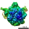 1127MC 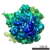 1128MC 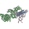 1zn0C C: citing same article ( M: map data used to model this data |
|---|---|
| Similar structure data |
-Links
 PDBj
PDBj


- Assembly
| Deposited unit | 
|
|---|---|
| 1 |
|
-Components
| #1: RNA chain | Mass: 18952.248 Da / Num. of mol.: 1 / Fragment: fragment of large subunit rRNA helix 69-71 / Source method: isolated from a natural source / Source: (natural) |
|---|---|
| #2: RNA chain | Mass: 12988.728 Da / Num. of mol.: 1 / Fragment: fragment of small subunit rRNA helix 44 / Source method: isolated from a natural source / Source: (natural) |
| #3: Protein | Mass: 20671.621 Da / Num. of mol.: 1 / Source method: isolated from a natural source / Source: (natural) |
| #4: Protein | Mass: 10828.608 Da / Num. of mol.: 1 / Source method: isolated from a natural source / Source: (natural) |
-Experimental details
-Experiment
| Experiment | Method: ELECTRON MICROSCOPY |
|---|---|
| EM experiment | Aggregation state: PARTICLE / 3D reconstruction method: single particle reconstruction |
- Sample preparation
| Component | Name: 70S ribosome / Type: RIBOSOME |
|---|---|
| Buffer solution | Name: Polymix buffer / pH: 7.5 / Details: Polymix buffer |
| Specimen | Conc.: 0.032 mg/ml / Embedding applied: NO / Shadowing applied: NO / Staining applied: NO / Vitrification applied: YES |
| Specimen support | Details: Quantifoil holey carbon film grids |
| Vitrification | Instrument: HOMEMADE PLUNGER / Cryogen name: ETHANE / Details: Rapid-freezing in liquid ethane |
- Electron microscopy imaging
| Experimental equipment |  Model: Tecnai F20 / Image courtesy: FEI Company |
|---|---|
| Microscopy | Model: FEI TECNAI F20 / Date: Feb 1, 2003 |
| Electron gun | Electron source:  FIELD EMISSION GUN / Accelerating voltage: 200 kV / Illumination mode: FLOOD BEAM FIELD EMISSION GUN / Accelerating voltage: 200 kV / Illumination mode: FLOOD BEAM |
| Electron lens | Mode: BRIGHT FIELD / Nominal magnification: 50000 X / Calibrated magnification: 49700 X / Nominal defocus max: -3.8 nm / Nominal defocus min: -1.4 nm |
| Specimen holder | Temperature: 93 K / Tilt angle max: 0 ° / Tilt angle min: 0 ° |
| Image recording | Electron dose: 15 e/Å2 / Film or detector model: KODAK SO-163 FILM |
- Processing
| CTF correction | Details: CTF correction of 3D maps by Wiener filtration | ||||||||||||||||||||||||||||
|---|---|---|---|---|---|---|---|---|---|---|---|---|---|---|---|---|---|---|---|---|---|---|---|---|---|---|---|---|---|
| Symmetry | Point symmetry: C1 (asymmetric) | ||||||||||||||||||||||||||||
| 3D reconstruction | Method: 3D projection matching; conjugate gradient with regularization Resolution: 14.1 Å / Num. of particles: 37379 / Actual pixel size: 2.82 Å / Magnification calibration: TMV / Details: SPIDER package / Symmetry type: POINT | ||||||||||||||||||||||||||||
| Atomic model building | Protocol: OTHER / Space: REAL / Details: METHOD--Manual fitting in O | ||||||||||||||||||||||||||||
| Atomic model building |
| ||||||||||||||||||||||||||||
| Refinement step | Cycle: LAST
|

