 Movie
Movie Controller

[English] 日本語
 Yorodumi
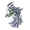
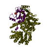
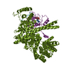
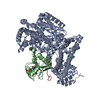
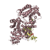
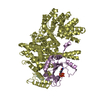
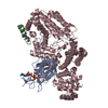
- PDB-1ct9: CRYSTAL STRUCTURE OF ASPARAGINE SYNTHETASE B FROM ESCHERICHIA COLI -
+ Open data

- Basic information
| Entry | Database: PDB / ID: 1ct9 | ||||||
|---|---|---|---|---|---|---|---|
| Title | CRYSTAL STRUCTURE OF ASPARAGINE SYNTHETASE B FROM ESCHERICHIA COLI | ||||||
 Components Components | ASPARAGINE SYNTHETASE B | ||||||
 Keywords Keywords | LIGASE / AMIDOTRANSFERASE / SUBSTRATE CHANNELING / ASPARAGINE BIOSYNTHESIS | ||||||
| Function / homology |  Function and homology information Function and homology informationaspartate-ammonia ligase activity / asparagine synthase (glutamine-hydrolysing) / L-asparagine biosynthetic process / asparagine synthase (glutamine-hydrolyzing) activity / : / amino acid catabolic process / L-glutamine metabolic process / amino acid binding / amino acid biosynthetic process / protein homodimerization activity ...aspartate-ammonia ligase activity / asparagine synthase (glutamine-hydrolysing) / L-asparagine biosynthetic process / asparagine synthase (glutamine-hydrolyzing) activity / : / amino acid catabolic process / L-glutamine metabolic process / amino acid binding / amino acid biosynthetic process / protein homodimerization activity / ATP binding / identical protein binding / cytoplasm / cytosol Similarity search - Function | ||||||
| Biological species |  | ||||||
| Method |  X-RAY DIFFRACTION / SYNCHROTRON / Resolution: 2 Å X-RAY DIFFRACTION / SYNCHROTRON / Resolution: 2 Å | ||||||
 Authors Authors | Larsen, T.M. / Boehlein, S.K. / Schuster, S.M. / Richards, N.G.J. / Thoden, J.B. / Holden, H.M. / Rayment, I. | ||||||
 Citation Citation | Journal: Biochemistry / Year: 1999 Title: Three-dimensional structure of Escherichia coli asparagine synthetase B: a short journey from substrate to product. Authors: Larsen, T.M. / Boehlein, S.K. / Schuster, S.M. / Richards, N.G. / Thoden, J.B. / Holden, H.M. / Rayment, I. | ||||||
| History |
|
- Structure visualization
| Structure viewer | Molecule: MolmilJmol/JSmol |
|---|
- Downloads & links
-Download
| PDBx/mmCIF format | 1ct9.cif.gz | 428.2 KB | Display | PDBx/mmCIF format |
|---|---|---|---|---|
| PDB format | pdb1ct9.ent.gz | 344.1 KB | Display | PDB format |
| PDBx/mmJSON format | 1ct9.json.gz | Tree view | PDBx/mmJSON format | |
| Others |  Other downloads Other downloads |
-Validation report
| Arichive directory | https://data.pdbj.org/pub/pdb/validation_reports/ct/1ct9ftp://data.pdbj.org/pub/pdb/validation_reports/ct/1ct9 | HTTPS FTP |
|---|
-Related structure data
| Similar structure data |
|---|


-Links
 PDBj
PDBj


- Assembly
| Deposited unit | 
| ||||||||
|---|---|---|---|---|---|---|---|---|---|
| 1 | 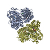
| ||||||||
| 2 | 
| ||||||||
| Unit cell |
|
-Components
-Protein , 1 types, 4 molecules ABCD
| #1: Protein | Mass: 62575.672 Da / Num. of mol.: 4 / Mutation: C1A Source method: isolated from a genetically manipulated source Source: (gene. exp.) References: UniProt: ASNB_ECOLI, UniProt: P22106*PLUS, asparagine synthase (glutamine-hydrolysing) |
|---|
-Non-polymers , 5 types, 1074 molecules 


| #2: Chemical | ChemComp-IUM /  Mass: 270.028 Da / Num. of mol.: 16 / Source method: obtained synthetically / Formula: O2U Mass: 270.028 Da / Num. of mol.: 16 / Source method: obtained synthetically / Formula: O2U#3: Chemical | ChemComp-CL /  Mass: 35.453 Da / Num. of mol.: 4 / Source method: obtained synthetically / Formula: Cl Mass: 35.453 Da / Num. of mol.: 4 / Source method: obtained synthetically / Formula: Cl#4: Chemical | ChemComp-AMP /  Mass: 347.221 Da / Num. of mol.: 4 / Source method: obtained synthetically / Formula: C10H14N5O7P / Comment: AMP*YM Mass: 347.221 Da / Num. of mol.: 4 / Source method: obtained synthetically / Formula: C10H14N5O7P / Comment: AMP*YM#5: Chemical | ChemComp-GLN /  Type: L-peptide linking / Mass: 146.144 Da / Num. of mol.: 4 / Source method: obtained synthetically / Formula: C5H10N2O3 Type: L-peptide linking / Mass: 146.144 Da / Num. of mol.: 4 / Source method: obtained synthetically / Formula: C5H10N2O3#6: Water | ChemComp-HOH / | Mass: 18.015 Da / Num. of mol.: 1046 / Source method: isolated from a natural source / Formula: H2O |
|---|
-Experimental details
-Experiment
| Experiment | Method: X-RAY DIFFRACTION / Number of used crystals: 1 |
|---|
- Sample preparation
| Crystal | Density Matthews: 2.59 Å3/Da / Density % sol: 52.57 % | ||||||||||||||||||||||||||||||||||||||||||||||||||||||
|---|---|---|---|---|---|---|---|---|---|---|---|---|---|---|---|---|---|---|---|---|---|---|---|---|---|---|---|---|---|---|---|---|---|---|---|---|---|---|---|---|---|---|---|---|---|---|---|---|---|---|---|---|---|---|---|
| Crystal grow | Temperature: 273 K / Method: vapor diffusion, hanging drop / pH: 7 Details: PEG 8000, HEPES, MAGNESIUM CHLORIDE, L-GLUTAMINE, ADENOSINE 5'-MONOPHOSPHATE, SODIUM CHLORIDE, pH 7.0, VAPOR DIFFUSION, HANGING DROP, temperature 273K | ||||||||||||||||||||||||||||||||||||||||||||||||||||||
| Crystal grow | *PLUS Temperature: 4 ℃ | ||||||||||||||||||||||||||||||||||||||||||||||||||||||
| Components of the solutions | *PLUS
|
-Data collection
| Diffraction | Mean temperature: 100 K |
|---|---|
| Diffraction source | Source: SYNCHROTRON / Site: APS  / Beamline: 19-ID / Wavelength: 0.7433 / Beamline: 19-ID / Wavelength: 0.7433 |
| Detector | Type: OTHER / Detector: CCD / Date: Nov 11, 1998 |
| Radiation | Protocol: SINGLE WAVELENGTH / Monochromatic (M) / Laue (L): M / Scattering type: x-ray |
| Radiation wavelength | Wavelength: 0.7433 Å / Relative weight: 1 |
| Reflection | Resolution: 1.9→30 Å / Num. all: 200469 / Num. obs: 200469 / % possible obs: 98 % / Observed criterion σ(F): 0 / Observed criterion σ(I): 0 / Redundancy: 5.4 % / Rmerge(I) obs: 0.063 / Net I/σ(I): 16.6 |
| Reflection shell | Resolution: 1.9→1.97 Å / Redundancy: 3.2 % / Rmerge(I) obs: 0.331 / % possible all: 93.6 |
| Reflection | *PLUS Num. measured all: 1085969 |
| Reflection shell | *PLUS % possible obs: 93.6 % |
- Processing
| Software |
| ||||||||||||||||||||||||||||||
|---|---|---|---|---|---|---|---|---|---|---|---|---|---|---|---|---|---|---|---|---|---|---|---|---|---|---|---|---|---|---|---|
| Refinement | Resolution: 2→30 Å / σ(F): 0 / σ(I): 0 / Stereochemistry target values: ENGH & HUBER
| ||||||||||||||||||||||||||||||
| Refinement step | Cycle: LAST / Resolution: 2→30 Å
| ||||||||||||||||||||||||||||||
| Refine LS restraints |
| ||||||||||||||||||||||||||||||
| Software | *PLUS Name: TNT / Classification: refinement | ||||||||||||||||||||||||||||||
| Refinement | *PLUS | ||||||||||||||||||||||||||||||
| Solvent computation | *PLUS | ||||||||||||||||||||||||||||||
| Displacement parameters | *PLUS | ||||||||||||||||||||||||||||||
| Refine LS restraints | *PLUS
|
