 Movie
Movie Controller

[English] 日本語
 Yorodumi
- PDB-1pjq: Structure and function of CysG, the multifunctional methyltransfe... -
+ Open data

- Basic information
| Entry | Database: PDB / ID: 1pjq | ||||||
|---|---|---|---|---|---|---|---|
| Title | Structure and function of CysG, the multifunctional methyltransferase/dehydrogenase/ferrochelatase for siroheme synthesis | ||||||
 Components Components | Siroheme synthase | ||||||
 Keywords Keywords | TRANSFERASE/OXIDOREDUCTASE/LYASE / Rossmann fold / nucleotide binding motif / SAM / NAD / phosphoserine / TRANSFERASE-OXIDOREDUCTASE-LYASE COMPLEX | ||||||
| Function / homology |  Function and homology information Function and homology informationprecorrin-2 dehydrogenase / precorrin-2 dehydrogenase activity / uroporphyrinogen-III C-methyltransferase / uroporphyrin-III C-methyltransferase activity / sirohydrochlorin ferrochelatase / sirohydrochlorin ferrochelatase activity / siroheme biosynthetic process / cobalamin biosynthetic process / NAD binding / methylation Similarity search - Function | ||||||
| Biological species |  Salmonella typhimurium (bacteria) Salmonella typhimurium (bacteria) | ||||||
| Method |  X-RAY DIFFRACTION / SYNCHROTRON / MAD / Resolution: 2.21 Å X-RAY DIFFRACTION / SYNCHROTRON / MAD / Resolution: 2.21 Å | ||||||
 Authors Authors | Stroupe, M.E. / Leech, H.K. / Daniels, D.S. / Warren, M.J. / Getzoff, E.D. | ||||||
 Citation Citation | Journal: Nat.Struct.Biol. / Year: 2003 Title: CysG structure reveals tetrapyrrole-binding features and novel regulation of siroheme biosynthesis. Authors: Stroupe, M.E. / Leech, H.K. / Daniels, D.S. / Warren, M.J. / Getzoff, E.D. | ||||||
| History |
|
- Structure visualization
| Structure viewer | Molecule: MolmilJmol/JSmol |
|---|
- Downloads & links
-Download
| PDBx/mmCIF format | 1pjq.cif.gz | 196 KB | Display | PDBx/mmCIF format |
|---|---|---|---|---|
| PDB format | pdb1pjq.ent.gz | 156.7 KB | Display | PDB format |
| PDBx/mmJSON format | 1pjq.json.gz | Tree view | PDBx/mmJSON format | |
| Others |  Other downloads Other downloads |
-Validation report
| Arichive directory | https://data.pdbj.org/pub/pdb/validation_reports/pj/1pjqftp://data.pdbj.org/pub/pdb/validation_reports/pj/1pjq | HTTPS FTP |
|---|
-Related structure data


-Links
 PDBj
PDBj- Assembly
| Deposited unit | 
| ||||||||
|---|---|---|---|---|---|---|---|---|---|
| 1 |
| ||||||||
| Unit cell |
| ||||||||
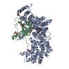
| Details | The asymmetric unit contains a single homodimer. Each of CysG's modules (CysGB, residues 1-212, and CysGA, residues 213-457) are homodimers. Each subunit contains half of CysGB and half of CysGA, each of which is related to its dimer partner by a unique non-crystallographic two-fold symmetry axis. CysG has two distinct non-crystallographic two-fold symmetry axes, one for each homodimeric module. |
-Components
| #1: Protein | Mass: 50286.477 Da / Num. of mol.: 2 Source method: isolated from a genetically manipulated source Details: Residues 1-212 (CysGB) are a dehydrogenase/ferrochelatase, Residues 213-457 (CysGA) are a bismethyltransferase Source: (gene. exp.) Salmonella typhimurium (bacteria) / Gene: CYSG OR STM3477 / Plasmid: PJYW632-C483GSIRHP / Production host: Salmonella enterica (bacteria) / Strain (production host): SA2853References: UniProt: P25924, uroporphyrinogen-III C-methyltransferase, Oxidoreductases, EC: 4.99.1.- #2: Chemical |   Mass: 59.044 Da / Num. of mol.: 3 / Source method: obtained synthetically / Formula: C2H3O2 Mass: 59.044 Da / Num. of mol.: 3 / Source method: obtained synthetically / Formula: C2H3O2#3: Chemical | ChemComp-PGE / |   Mass: 150.173 Da / Num. of mol.: 1 / Source method: obtained synthetically / Formula: C6H14O4 Mass: 150.173 Da / Num. of mol.: 1 / Source method: obtained synthetically / Formula: C6H14O4#4: Chemical | ChemComp-SAH / |   Mass: 384.411 Da / Num. of mol.: 1 / Source method: obtained synthetically / Formula: C14H20N6O5S Mass: 384.411 Da / Num. of mol.: 1 / Source method: obtained synthetically / Formula: C14H20N6O5S#5: Water | ChemComp-HOH / |  Mass: 18.015 Da / Num. of mol.: 326 / Source method: isolated from a natural source / Formula: H2O Mass: 18.015 Da / Num. of mol.: 326 / Source method: isolated from a natural source / Formula: H2OHas protein modification | Y | |
|---|
-Experimental details
-Experiment
| Experiment | Method: X-RAY DIFFRACTION / Number of used crystals: 1 |
|---|
- Sample preparation
| Crystal | Density Matthews: 2.4 Å3/Da / Density % sol: 48.71 % | ||||||||||||||||||||||||||||||||||||||||||
|---|---|---|---|---|---|---|---|---|---|---|---|---|---|---|---|---|---|---|---|---|---|---|---|---|---|---|---|---|---|---|---|---|---|---|---|---|---|---|---|---|---|---|---|
| Crystal grow | Temperature: 293 K / Method: vapor diffusion, hanging drop / pH: 4.8 Details: PEG200, acetate, sodium chloride, 2-mercaptoethanol, pH 4.8, VAPOR DIFFUSION, HANGING DROP, temperature 293K | ||||||||||||||||||||||||||||||||||||||||||
| Crystal grow | *PLUS pH: 4.5 / Method: vapor diffusion | ||||||||||||||||||||||||||||||||||||||||||
| Components of the solutions | *PLUS
|
-Data collection
| Diffraction | Mean temperature: 100 K |
|---|---|
| Diffraction source | Source: SYNCHROTRON / Site: SSRL  / Beamline: BL9-1 / Wavelength: 0.98 Å / Beamline: BL9-1 / Wavelength: 0.98 Å |
| Detector | Type: MARRESEARCH / Detector: IMAGE PLATE / Date: Mar 10, 2000 Details: flat mirror (vertical focusing) single crystal (Si311) bent monochromator (horizontal focusing) |
| Radiation | Monochromator: Si(311) bent / Protocol: SINGLE WAVELENGTH / Monochromatic (M) / Laue (L): M / Scattering type: x-ray |
| Radiation wavelength | Wavelength: 0.98 Å / Relative weight: 1 |
| Reflection | Resolution: 2.21→19.97 Å / Num. obs: 48911 / % possible obs: 99.5 % / Observed criterion σ(F): 0 / Observed criterion σ(I): 0 / Redundancy: 3.5 % / Biso Wilson estimate: 24.3 Å2 / Rsym value: 0.057 / Net I/σ(I): 23 |
| Reflection shell | Resolution: 2.21→2.26 Å / Mean I/σ(I) obs: 3 / Num. unique all: 2348 / Rsym value: 0.453 / % possible all: 97.5 |
| Reflection | *PLUS Lowest resolution: 20 Å / Num. obs: 48472 / Rmerge(I) obs: 0.057 |
| Reflection shell | *PLUS % possible obs: 97.5 % / Rmerge(I) obs: 0.453 |
- Processing
| Software |
| ||||||||||||||||||||||||
|---|---|---|---|---|---|---|---|---|---|---|---|---|---|---|---|---|---|---|---|---|---|---|---|---|---|
| Refinement | Method to determine structure: MAD / Resolution: 2.21→19.97 Å / Rfactor Rfree error: 0.004 / Isotropic thermal model: RESTRAINED / Cross valid method: THROUGHOUT / σ(F): 0 / Stereochemistry target values: Engh & Huber
| ||||||||||||||||||||||||
| Solvent computation | Solvent model: FLAT MODEL / Bsol: 55.0306 Å2 / ksol: 0.332865 e/Å3 | ||||||||||||||||||||||||
| Displacement parameters | Biso mean: 40.2 Å2
| ||||||||||||||||||||||||
| Refine analyze |
| ||||||||||||||||||||||||
| Refinement step | Cycle: LAST / Resolution: 2.21→19.97 Å
| ||||||||||||||||||||||||
| Refine LS restraints |
| ||||||||||||||||||||||||
| LS refinement shell | Resolution: 2.21→2.34 Å / Rfactor Rfree error: 0.012 / Total num. of bins used: 6
| ||||||||||||||||||||||||
| Xplor file |
| ||||||||||||||||||||||||
| Refinement | *PLUS Lowest resolution: 20 Å / % reflection Rfree: 10 % / Rfactor Rfree: 0.275 / Rfactor Rwork: 0.236 | ||||||||||||||||||||||||
| Solvent computation | *PLUS | ||||||||||||||||||||||||
| Displacement parameters | *PLUS | ||||||||||||||||||||||||
| Refine LS restraints | *PLUS
|
