 Movie
Movie Controller

[English] 日本語
 Yorodumi
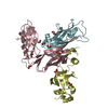
- PDB-1ze3: Crystal Structure of the Ternary Complex of FIMD (N-Terminal Doma... -
+ Open data

- Basic information
| Entry | Database: PDB / ID: 1ze3 | ||||||
|---|---|---|---|---|---|---|---|
| Title | Crystal Structure of the Ternary Complex of FIMD (N-Terminal Domain) with FIMC and the Pilin Domain of FIMH | ||||||
 Components Components |
| ||||||
 Keywords Keywords | CHAPERONE/STRUCTURAL/MEMBRANE PROTEIN / Usher / soluble domain / ternary complex with chaperone and pilus subunit / CHAPERONE-STRUCTURAL-MEMBRANE PROTEIN COMPLEX | ||||||
| Function / homology |  Function and homology information Function and homology informationfimbrial usher porin activity / pilus assembly / pilus tip / mechanosensory behavior / Attachment of bacteria to epithelial cells / cell adhesion involved in single-species biofilm formation / pilus / cell-substrate adhesion / D-mannose binding / host cell membrane ...fimbrial usher porin activity / pilus assembly / pilus tip / mechanosensory behavior / Attachment of bacteria to epithelial cells / cell adhesion involved in single-species biofilm formation / pilus / cell-substrate adhesion / D-mannose binding / host cell membrane / protein folding chaperone / cell outer membrane / cell wall organization / outer membrane-bounded periplasmic space / protein folding / cell adhesion Similarity search - Function | ||||||
| Biological species |  | ||||||
| Method |  X-RAY DIFFRACTION / SYNCHROTRON / MOLECULAR REPLACEMENT / Resolution: 1.84 Å X-RAY DIFFRACTION / SYNCHROTRON / MOLECULAR REPLACEMENT / Resolution: 1.84 Å | ||||||
 Authors Authors | Nishiyama, M. / Horst, R. / Eidam, O. / Herrmann, T. / Ignatov, O. / Vetsch, M. / Bettendorff, P. / Jelesarov, I. / Grutter, M.G. / Wuthrich, K. ...Nishiyama, M. / Horst, R. / Eidam, O. / Herrmann, T. / Ignatov, O. / Vetsch, M. / Bettendorff, P. / Jelesarov, I. / Grutter, M.G. / Wuthrich, K. / Glockshuber, R. / Capitani, G. | ||||||
 Citation Citation | Journal: Embo J. / Year: 2005 Title: Structural basis of chaperone-subunit complex recognition by the type 1 pilus assembly platform FimD. Authors: Nishiyama, M. / Horst, R. / Eidam, O. / Herrmann, T. / Ignatov, O. / Vetsch, M. / Bettendorff, P. / Jelesarov, I. / Glockshuber, R. / Capitani, G. | ||||||
| History |
|
- Structure visualization
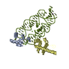
| Structure viewer | Molecule: MolmilJmol/JSmol |
|---|
- Downloads & links
-Download
| PDBx/mmCIF format | 1ze3.cif.gz | 111.9 KB | Display | PDBx/mmCIF format |
|---|---|---|---|---|
| PDB format | pdb1ze3.ent.gz | 84 KB | Display | PDB format |
| PDBx/mmJSON format | 1ze3.json.gz | Tree view | PDBx/mmJSON format | |
| Others |  Other downloads Other downloads |
-Validation report
| Arichive directory | https://data.pdbj.org/pub/pdb/validation_reports/ze/1ze3ftp://data.pdbj.org/pub/pdb/validation_reports/ze/1ze3 | HTTPS FTP |
|---|
-Related structure data
| Related structure data | 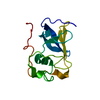 1zdvC  1zdxC  1qunS S: Starting model for refinement C: citing same article ( |
|---|---|
| Similar structure data |


-Links
 PDBj
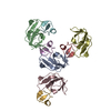
PDBj- Assembly
| Deposited unit | 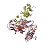
| ||||||||
|---|---|---|---|---|---|---|---|---|---|
| 1 |
| ||||||||
| Unit cell |
|
-Components
| #1: Protein | Mass: 22754.031 Da / Num. of mol.: 1 / Fragment: FimC Source method: isolated from a genetically manipulated source Source: (gene. exp.) | ||||
|---|---|---|---|---|---|
| #2: Protein | Mass: 12283.583 Da / Num. of mol.: 1 / Fragment: FimH pilin domain Source method: isolated from a genetically manipulated source Source: (gene. exp.) | ||||
| #3: Protein | Mass: 13664.263 Da / Num. of mol.: 1 / Fragment: FimD N-terminal domain Source method: isolated from a genetically manipulated source Source: (gene. exp.) | ||||
| #4: Chemical | ChemComp-EDO /   Mass: 62.068 Da / Num. of mol.: 12 / Source method: obtained synthetically / Formula: C2H6O2 Mass: 62.068 Da / Num. of mol.: 12 / Source method: obtained synthetically / Formula: C2H6O2#5: Water | ChemComp-HOH / |  Mass: 18.015 Da / Num. of mol.: 508 / Source method: isolated from a natural source / Formula: H2O Mass: 18.015 Da / Num. of mol.: 508 / Source method: isolated from a natural source / Formula: H2OHas protein modification | Y | |
-Experimental details
-Experiment
| Experiment | Method: X-RAY DIFFRACTION / Number of used crystals: 1 |
|---|
- Sample preparation
| Crystal | Density Matthews: 2.6 Å3/Da / Density % sol: 44 % |
|---|---|
| Crystal grow | Temperature: 293 K / Method: vapor diffusion, sitting drop / pH: 6.5 Details: PEG 6000, MES buffer, pH 6.5, VAPOR DIFFUSION, SITTING DROP, temperature 293K |
-Data collection
| Diffraction | Mean temperature: 100 K |
|---|---|
| Diffraction source | Source: SYNCHROTRON / Site: SLS  / Beamline: X06SA / Wavelength: 0.9 Å / Beamline: X06SA / Wavelength: 0.9 Å |
| Detector | Type: MARRESEARCH / Detector: CCD / Date: Jul 15, 2004 / Details: DYNAMICALLY BENDABLE MIRROR |
| Radiation | Monochromator: SI(111) / Protocol: SINGLE WAVELENGTH / Monochromatic (M) / Laue (L): M / Scattering type: x-ray |
| Radiation wavelength | Wavelength: 0.9 Å / Relative weight: 1 |
| Reflection | Resolution: 1.843→33.615 Å / Num. obs: 44185 / % possible obs: 99.9 % / Observed criterion σ(I): -3 / Redundancy: 6.4 % / Biso Wilson estimate: 15.8 Å2 / Rsym value: 0.088 / Net I/σ(I): 15 |
| Reflection shell | Resolution: 1.84→1.91 Å / Mean I/σ(I) obs: 3.1 / Rsym value: 0.363 / % possible all: 99.5 |
- Processing
| Software |
| ||||||||||||||||
|---|---|---|---|---|---|---|---|---|---|---|---|---|---|---|---|---|---|
| Refinement | Method to determine structure: MOLECULAR REPLACEMENT Starting model: MODEL DERIVED FROM PDB ENTRY 1QUN Resolution: 1.84→33.7 Å / Isotropic thermal model: ISOTROPIC / Cross valid method: THROUGHOUT / σ(F): 0 / Stereochemistry target values: Engh & Huber
| ||||||||||||||||
| Displacement parameters | Biso mean: 18.8 Å2
| ||||||||||||||||
| Refine analyze |
| ||||||||||||||||
| Refinement step | Cycle: LAST / Resolution: 1.84→33.7 Å
| ||||||||||||||||
| Refine LS restraints |
| ||||||||||||||||
| LS refinement shell | Resolution: 1.84→1.91 Å
|
