 Movie
Movie Controller

[English] 日本語
 Yorodumi
- PDB-4xco: Signal-sequence induced conformational changes in the signal reco... -
+ Open data

- Basic information
| Entry | Database: PDB / ID: 4xco | ||||||
|---|---|---|---|---|---|---|---|
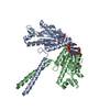
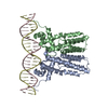
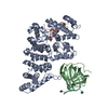
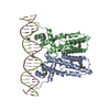
| Title | Signal-sequence induced conformational changes in the signal recognition particle | ||||||
 Components Components |
| ||||||
 Keywords Keywords | RNA BINDING PROTEIN / SIGNAL RECOGNITION PARTICLE / SIGNAL SEQUENCE / RECOMBINANT FUSION PROTEIN / RNA STRUCTURE | ||||||
| Function / homology |  Function and homology information Function and homology informationsignal recognition particle / signal-recognition-particle GTPase / 7S RNA binding / SRP-dependent cotranslational protein targeting to membrane / molecular adaptor activity / GTPase activity / GTP binding Similarity search - Function | ||||||
| Biological species |   Methanocaldococcus jannaschii (archaea) Methanocaldococcus jannaschii (archaea) | ||||||
| Method |  X-RAY DIFFRACTION / SYNCHROTRON / MOLECULAR REPLACEMENT / Resolution: 2.9 Å X-RAY DIFFRACTION / SYNCHROTRON / MOLECULAR REPLACEMENT / Resolution: 2.9 Å | ||||||
 Authors Authors | Hainzl, T. / Sauer-Eriksson, A.E. | ||||||
| Funding support |  Sweden, 1items Sweden, 1items
| ||||||
 Citation Citation | Journal: Nat Commun / Year: 2015 Title: Signal-sequence induced conformational changes in the signal recognition particle. Authors: Hainzl, T. / Sauer-Eriksson, A.E. #1: Journal: Nat. Struct. Mol. Biol. / Year: 2011Title: Structural basis of signal-sequence recognition by the signal recognition particle. Authors: Hainzl, T. / Huang, S. / Merilainen, G. / Brannstrom, K. / Sauer-Eriksson, A.E. | ||||||
| History |
|
- Structure visualization
| Structure viewer | Molecule: MolmilJmol/JSmol |
|---|
- Downloads & links
-Download
| PDBx/mmCIF format | 4xco.cif.gz | 334.6 KB | Display | PDBx/mmCIF format |
|---|---|---|---|---|
| PDB format | pdb4xco.ent.gz | 265.1 KB | Display | PDB format |
| PDBx/mmJSON format | 4xco.json.gz | Tree view | PDBx/mmJSON format | |
| Others |  Other downloads Other downloads |
-Validation report
| Arichive directory | https://data.pdbj.org/pub/pdb/validation_reports/xc/4xcoftp://data.pdbj.org/pub/pdb/validation_reports/xc/4xco | HTTPS FTP |
|---|
-Related structure data
| Related structure data |  3ndbS S: Starting model for refinement |
|---|---|
| Similar structure data |


-Links
 PDBj
PDBj


- Assembly
| Deposited unit | 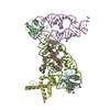
| ||||||||
|---|---|---|---|---|---|---|---|---|---|
| 1 | 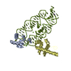
| ||||||||
| 2 | 
| ||||||||
| Unit cell |
|
-Components
-RNA chain , 1 types, 2 molecules ME
| #1: RNA chain | Mass: 31162.604 Da / Num. of mol.: 2 Source method: isolated from a genetically manipulated source Source: (gene. exp.) Methanocaldococcus jannaschii (archaea)Production host: synthetic construct (others) / References: GenBank: 6626255 |
|---|
-Signal recognition particle ... , 2 types, 4 molecules ABCD
| #2: Protein | Mass: 10376.414 Da / Num. of mol.: 2 Source method: isolated from a genetically manipulated source Source: (gene. exp.) Methanocaldococcus jannaschii (archaea)Gene: srp19, MJ1034 / Plasmid: pET3a / Production host:  #3: Protein | Mass: 19206.736 Da / Num. of mol.: 2 Source method: isolated from a genetically manipulated source Source: (gene. exp.) Methanocaldococcus jannaschii (archaea)Gene: srp54, MJ0101, mj0101, srp54 / Plasmid: pNZ8048 / Production host: Lactococcus lactis (lactic acid bacteria) / References: UniProt: Q57565 |
|---|
-Non-polymers , 3 types, 85 molecules 


| #4: Chemical | ChemComp-MG /  Mass: 24.305 Da / Num. of mol.: 14 / Source method: obtained synthetically / Formula: Mg Mass: 24.305 Da / Num. of mol.: 14 / Source method: obtained synthetically / Formula: Mg#5: Chemical | ChemComp-NA / |  Mass: 22.990 Da / Num. of mol.: 1 / Source method: obtained synthetically / Formula: Na Mass: 22.990 Da / Num. of mol.: 1 / Source method: obtained synthetically / Formula: Na#6: Water | ChemComp-HOH / | Mass: 18.015 Da / Num. of mol.: 70 / Source method: isolated from a natural source / Formula: H2O |
|---|
-Experimental details
-Experiment
| Experiment | Method: X-RAY DIFFRACTION |
|---|
- Sample preparation
| Crystal | Density Matthews: 2.57 Å3/Da / Density % sol: 52.15 % |
|---|---|
| Crystal grow | Temperature: 291 K / Method: vapor diffusion, hanging drop / pH: 5 Details: The complex (3mg/ml) was mixed 1:1 with 35% 2-Methyl-2,4-pentanediol (MPD), 200 mM NaCl, 80 mM MgCl2, 100 mM sodium acetate |
-Data collection
| Diffraction | Mean temperature: 100 K |
|---|---|
| Diffraction source | Source: SYNCHROTRON / Site: ESRF  / Beamline: ID23-1 / Wavelength: 0.984 Å / Beamline: ID23-1 / Wavelength: 0.984 Å |
| Detector | Type: DECTRIS PILATUS 6M-F / Detector: PIXEL / Date: Feb 12, 2014 |
| Radiation | Protocol: SINGLE WAVELENGTH / Monochromatic (M) / Laue (L): M / Scattering type: x-ray |
| Radiation wavelength | Wavelength: 0.984 Å / Relative weight: 1 |
| Reflection | Resolution: 2.9→48.025 Å / Num. obs: 28330 / % possible obs: 99.7 % / Redundancy: 6.5 % / Rmerge(I) obs: 0.073 / Net I/σ(I): 13.7 |
| Reflection shell | Resolution: 2.9→3.08 Å / Redundancy: 6.7 % / Rmerge(I) obs: 0.899 / Mean I/σ(I) obs: 1.9 / % possible all: 99.7 |
- Processing
| Software |
| |||||||||||||||||||||||||||||||||||||||||||||||||||||||||||||||||||||||||||||
|---|---|---|---|---|---|---|---|---|---|---|---|---|---|---|---|---|---|---|---|---|---|---|---|---|---|---|---|---|---|---|---|---|---|---|---|---|---|---|---|---|---|---|---|---|---|---|---|---|---|---|---|---|---|---|---|---|---|---|---|---|---|---|---|---|---|---|---|---|---|---|---|---|---|---|---|---|---|---|
| Refinement | Method to determine structure: MOLECULAR REPLACEMENT Starting model: 3ndb Resolution: 2.9→48.025 Å / SU ML: 0.37 / Cross valid method: FREE R-VALUE / σ(F): 1.34 / Phase error: 28.62 / Stereochemistry target values: ML
| |||||||||||||||||||||||||||||||||||||||||||||||||||||||||||||||||||||||||||||
| Solvent computation | Shrinkage radii: 0.9 Å / VDW probe radii: 1.11 Å / Solvent model: FLAT BULK SOLVENT MODEL | |||||||||||||||||||||||||||||||||||||||||||||||||||||||||||||||||||||||||||||
| Refinement step | Cycle: LAST / Resolution: 2.9→48.025 Å
| |||||||||||||||||||||||||||||||||||||||||||||||||||||||||||||||||||||||||||||
| Refine LS restraints |
| |||||||||||||||||||||||||||||||||||||||||||||||||||||||||||||||||||||||||||||
| LS refinement shell |
|
