 Movie
Movie Controller

[English] 日本語
 Yorodumi
- PDB-3ndb: Crystal structure of a signal sequence bound to the signal recogn... -
+ Open data

- Basic information
| Entry | Database: PDB / ID: 3ndb | |||||||||
|---|---|---|---|---|---|---|---|---|---|---|
| Title | Crystal structure of a signal sequence bound to the signal recognition particle | |||||||||
 Components Components |
| |||||||||
 Keywords Keywords | SIGNALING PROTEIN/RNA / protein-RNA complex / signal recognition particle / signal sequence / ribonucleoprotein / SIGNALING PROTEIN-RNA complex | |||||||||
| Function / homology |  Function and homology information Function and homology informationsignal recognition particle / signal-recognition-particle GTPase / 7S RNA binding / SRP-dependent cotranslational protein targeting to membrane / molecular adaptor activity / GTPase activity / GTP binding Similarity search - Function | |||||||||
| Biological species |   Methanocaldococcus jannaschii (archaea)Methanocaldococcus jannaschii DSM 2661 (archaea) Methanocaldococcus jannaschii (archaea)Methanocaldococcus jannaschii DSM 2661 (archaea) | |||||||||
| Method |  X-RAY DIFFRACTION / SYNCHROTRON / MOLECULAR REPLACEMENT / Resolution: 3 Å X-RAY DIFFRACTION / SYNCHROTRON / MOLECULAR REPLACEMENT / Resolution: 3 Å | |||||||||
 Authors Authors | Hainzl, T. / Huang, S. / Sauer-Eriksson, E. | |||||||||
 Citation Citation | Journal: Nat.Struct.Mol.Biol. / Year: 2011 Title: Structural basis of signal-sequence recognition by the signal recognition particle. Authors: Hainzl, T. / Huang, S. / Merilainen, G. / Brannstrom, K. / Sauer-Eriksson, A.E. | |||||||||
| History |
|
- Structure visualization
| Structure viewer | Molecule: MolmilJmol/JSmol |
|---|
- Downloads & links
-Download
| PDBx/mmCIF format | 3ndb.cif.gz | 369.3 KB | Display | PDBx/mmCIF format |
|---|---|---|---|---|
| PDB format | pdb3ndb.ent.gz | 297 KB | Display | PDB format |
| PDBx/mmJSON format | 3ndb.json.gz | Tree view | PDBx/mmJSON format | |
| Others |  Other downloads Other downloads |
-Validation report
| Arichive directory | https://data.pdbj.org/pub/pdb/validation_reports/nd/3ndbftp://data.pdbj.org/pub/pdb/validation_reports/nd/3ndb | HTTPS FTP |
|---|
-Related structure data
| Related structure data |  2v3cS S: Starting model for refinement |
|---|---|
| Similar structure data |
-Links
 PDBj
PDBj


- Assembly
| Deposited unit | 
| ||||||||
|---|---|---|---|---|---|---|---|---|---|
| 1 | 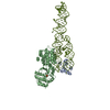
| ||||||||
| Unit cell |
|
-Components
| #1: Protein | Mass: 10376.414 Da / Num. of mol.: 1 Source method: isolated from a genetically manipulated source Source: (gene. exp.) Methanocaldococcus jannaschii (archaea)Gene: MJ1034, srp19 / Plasmid: pET3a / Production host:  |
|---|---|
| #2: Protein | Mass: 50014.723 Da / Num. of mol.: 1 Source method: isolated from a genetically manipulated source Source: (gene. exp.) Methanocaldococcus jannaschii (archaea)Gene: MJ0101, srp54 / Plasmid: pNZ8048 / Production host: Lactococcus lactis (lactic acid bacteria) / References: UniProt: Q57565 |
| #3: RNA chain | Mass: 44145.266 Da / Num. of mol.: 1 / Source method: obtained synthetically Source: (synth.) Methanocaldococcus jannaschii DSM 2661 (archaea)References: GenBank: L77117.1 |
| #4: Chemical | ChemComp-PO4 /   Mass: 94.971 Da / Num. of mol.: 1 / Source method: obtained synthetically / Formula: PO4 Mass: 94.971 Da / Num. of mol.: 1 / Source method: obtained synthetically / Formula: PO4 |
-Experimental details
-Experiment
| Experiment | Method: X-RAY DIFFRACTION / Number of used crystals: 1 |
|---|
- Sample preparation
| Crystal | Density Matthews: 3.32 Å3/Da / Density % sol: 62.9 % |
|---|---|
| Crystal grow | Temperature: 291 K / Method: evaporation / pH: 5.6 Details: 30% MPD, 200 mM NH4PO4 (pH 5.6), EVAPORATION, temperature 291K |
-Data collection
| Diffraction | Mean temperature: 100 K |
|---|---|
| Diffraction source | Source: SYNCHROTRON / Site: ESRF  / Beamline: ID23-1 / Wavelength: 1 Å / Beamline: ID23-1 / Wavelength: 1 Å |
| Detector | Type: ADSC QUANTUM 315r / Detector: CCD / Date: Feb 9, 2009 |
| Radiation | Monochromator: Silicon (1 1 1) channel-cut / Protocol: SINGLE WAVELENGTH / Monochromatic (M) / Laue (L): M / Scattering type: x-ray |
| Radiation wavelength | Wavelength: 1 Å / Relative weight: 1 |
| Reflection | Resolution: 3→63.8 Å / Num. obs: 27778 / % possible obs: 98.5 % / Observed criterion σ(F): 3 / Observed criterion σ(I): 9.2 / Redundancy: 4.8 % / Biso Wilson estimate: 14.1 Å2 / Rmerge(I) obs: 0.102 / Rsym value: 0.091 |
- Processing
| Software |
| ||||||||||||||||||||||||||||||||||||||||||||||||||||||||||||||||||||||||||||||||||||||||||||||||||||||||||||||||||||||||||||||||||||||||||||||||||||||||||||||||||||||||||||||||||||||||||||||||||||||||||||||||||||||||||||||||||||||||||||||||||||||||||||||||||||||||||||||||||||||||||||||||||||||||||||
|---|---|---|---|---|---|---|---|---|---|---|---|---|---|---|---|---|---|---|---|---|---|---|---|---|---|---|---|---|---|---|---|---|---|---|---|---|---|---|---|---|---|---|---|---|---|---|---|---|---|---|---|---|---|---|---|---|---|---|---|---|---|---|---|---|---|---|---|---|---|---|---|---|---|---|---|---|---|---|---|---|---|---|---|---|---|---|---|---|---|---|---|---|---|---|---|---|---|---|---|---|---|---|---|---|---|---|---|---|---|---|---|---|---|---|---|---|---|---|---|---|---|---|---|---|---|---|---|---|---|---|---|---|---|---|---|---|---|---|---|---|---|---|---|---|---|---|---|---|---|---|---|---|---|---|---|---|---|---|---|---|---|---|---|---|---|---|---|---|---|---|---|---|---|---|---|---|---|---|---|---|---|---|---|---|---|---|---|---|---|---|---|---|---|---|---|---|---|---|---|---|---|---|---|---|---|---|---|---|---|---|---|---|---|---|---|---|---|---|---|---|---|---|---|---|---|---|---|---|---|---|---|---|---|---|---|---|---|---|---|---|---|---|---|---|---|---|---|---|---|---|---|---|---|---|---|---|---|---|---|---|---|---|---|---|---|---|---|---|---|---|---|---|---|---|---|---|---|---|---|---|---|---|---|---|---|---|---|---|---|---|---|---|---|---|---|---|---|---|---|---|---|
| Refinement | Method to determine structure: MOLECULAR REPLACEMENT Starting model: PDB ENTRY 2V3C Resolution: 3→20 Å / Cor.coef. Fo:Fc: 0.932 / Cor.coef. Fo:Fc free: 0.894 / SU B: 46.48 / SU ML: 0.37 / Cross valid method: THROUGHOUT / σ(F): 0 / σ(I): 0 / ESU R Free: 0.42 / Stereochemistry target values: MAXIMUM LIKELIHOOD
| ||||||||||||||||||||||||||||||||||||||||||||||||||||||||||||||||||||||||||||||||||||||||||||||||||||||||||||||||||||||||||||||||||||||||||||||||||||||||||||||||||||||||||||||||||||||||||||||||||||||||||||||||||||||||||||||||||||||||||||||||||||||||||||||||||||||||||||||||||||||||||||||||||||||||||||
| Solvent computation | Ion probe radii: 0.8 Å / Shrinkage radii: 0.8 Å / VDW probe radii: 1.4 Å / Solvent model: BABINET MODEL WITH MASK | ||||||||||||||||||||||||||||||||||||||||||||||||||||||||||||||||||||||||||||||||||||||||||||||||||||||||||||||||||||||||||||||||||||||||||||||||||||||||||||||||||||||||||||||||||||||||||||||||||||||||||||||||||||||||||||||||||||||||||||||||||||||||||||||||||||||||||||||||||||||||||||||||||||||||||||
| Displacement parameters | Biso mean: 99.513 Å2
| ||||||||||||||||||||||||||||||||||||||||||||||||||||||||||||||||||||||||||||||||||||||||||||||||||||||||||||||||||||||||||||||||||||||||||||||||||||||||||||||||||||||||||||||||||||||||||||||||||||||||||||||||||||||||||||||||||||||||||||||||||||||||||||||||||||||||||||||||||||||||||||||||||||||||||||
| Refinement step | Cycle: LAST / Resolution: 3→20 Å
| ||||||||||||||||||||||||||||||||||||||||||||||||||||||||||||||||||||||||||||||||||||||||||||||||||||||||||||||||||||||||||||||||||||||||||||||||||||||||||||||||||||||||||||||||||||||||||||||||||||||||||||||||||||||||||||||||||||||||||||||||||||||||||||||||||||||||||||||||||||||||||||||||||||||||||||
| Refine LS restraints |
| ||||||||||||||||||||||||||||||||||||||||||||||||||||||||||||||||||||||||||||||||||||||||||||||||||||||||||||||||||||||||||||||||||||||||||||||||||||||||||||||||||||||||||||||||||||||||||||||||||||||||||||||||||||||||||||||||||||||||||||||||||||||||||||||||||||||||||||||||||||||||||||||||||||||||||||
| LS refinement shell | Resolution: 3→3.076 Å / Total num. of bins used: 20
| ||||||||||||||||||||||||||||||||||||||||||||||||||||||||||||||||||||||||||||||||||||||||||||||||||||||||||||||||||||||||||||||||||||||||||||||||||||||||||||||||||||||||||||||||||||||||||||||||||||||||||||||||||||||||||||||||||||||||||||||||||||||||||||||||||||||||||||||||||||||||||||||||||||||||||||
| Refinement TLS params. | Method: refined / Refine-ID: X-RAY DIFFRACTION
| ||||||||||||||||||||||||||||||||||||||||||||||||||||||||||||||||||||||||||||||||||||||||||||||||||||||||||||||||||||||||||||||||||||||||||||||||||||||||||||||||||||||||||||||||||||||||||||||||||||||||||||||||||||||||||||||||||||||||||||||||||||||||||||||||||||||||||||||||||||||||||||||||||||||||||||
| Refinement TLS group |
|
