 Movie
Movie Controller

[English] 日本語
 Yorodumi
- PDB-1yts: A LIGAND-INDUCED CONFORMATIONAL CHANGE IN THE YERSINIA PROTEIN TY... -
+ Open data

- Basic information
| Entry | Database: PDB / ID: 1yts | ||||||
|---|---|---|---|---|---|---|---|
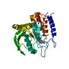
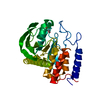
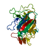
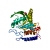
| Title | A LIGAND-INDUCED CONFORMATIONAL CHANGE IN THE YERSINIA PROTEIN TYROSINE PHOSPHATASE | ||||||
 Components Components | YERSINIA PROTEIN TYROSINE PHOSPHATASE | ||||||
 Keywords Keywords | HYDROLASE / PROTEIN TYROSINE PHOSPHATASE | ||||||
| Function / homology |  Function and homology information Function and homology informationprotein-tyrosine-phosphatase / protein tyrosine phosphatase activity / extracellular region Similarity search - Function | ||||||
| Biological species |  Yersinia enterocolitica (bacteria) Yersinia enterocolitica (bacteria) | ||||||
| Method |  X-RAY DIFFRACTION / Resolution: 2.5 Å X-RAY DIFFRACTION / Resolution: 2.5 Å | ||||||
 Authors Authors | Schubert, H.L. / Stuckey, J.A. / Fauman, E.B. / Dixon, J.E. / Saper, M.A. | ||||||
 Citation Citation | Journal: Protein Sci. / Year: 1995 Title: A ligand-induced conformational change in the Yersinia protein tyrosine phosphatase. Authors: Schubert, H.L. / Fauman, E.B. / Stuckey, J.A. / Dixon, J.E. / Saper, M.A. #1: Journal: Proc.Natl.Acad.Sci.USA / Year: 1994Title: Dissecting the Catalytic Mechanism of Protein-Tyrosine Phosphatases Authors: Zhang, Z.-Y. / Wang, Y. / Dixon, J.E. #2: Journal: Nature / Year: 1994Title: Crystal Structure of Yersinia Protein Tyrosine Phosphatase at 2.5 Angstroms and the Complex with Tungstate Authors: Stuckey, J.A. / Schubert, H.L. / Fauman, E.B. / Zhang, Z.-Y. / Dixon, J.E. / Saper, M.A. #3: Journal: J.Biol.Chem. / Year: 1992Title: Expression, Purification, and Physicochemical Characterization of a Recombinant Yersinia Protein Tyrosine Phosphatase Authors: Zhang, Z.-Y. / Clemens, J.C. / Schubert, H.L. / Stuckey, J.A. / Fischer, M.W.F. / Hume, D.M. / Saper, M.A. / Dixon, J.E. | ||||||
| History |
|
- Structure visualization
| Structure viewer | Molecule: MolmilJmol/JSmol |
|---|
- Downloads & links
-Download
| PDBx/mmCIF format | 1yts.cif.gz | 67.7 KB | Display | PDBx/mmCIF format |
|---|---|---|---|---|
| PDB format | pdb1yts.ent.gz | 49.9 KB | Display | PDB format |
| PDBx/mmJSON format | 1yts.json.gz | Tree view | PDBx/mmJSON format | |
| Others |  Other downloads Other downloads |
-Validation report
| Arichive directory | https://data.pdbj.org/pub/pdb/validation_reports/yt/1ytsftp://data.pdbj.org/pub/pdb/validation_reports/yt/1yts | HTTPS FTP |
|---|
-Related structure data
| Similar structure data |
|---|


-Links
 PDBj
PDBj


- Assembly
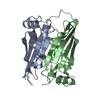
| Deposited unit | 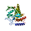
| ||||||||
|---|---|---|---|---|---|---|---|---|---|
| 1 |
| ||||||||
| Unit cell |
|
-Components
| #1: Protein | Mass: 30631.664 Da / Num. of mol.: 1 Mutation: CYS 235 REPLACED BY ARG, CYS 403 REPLACED BY SER, C235R, C403S Source method: isolated from a genetically manipulated source Source: (gene. exp.) Yersinia enterocolitica (bacteria) / Strain: W22703 / Gene: YOP51 / Plasmid: PT7-7 / Gene (production host): YOP51 / Production host: | ||||
|---|---|---|---|---|---|
| #2: Chemical |   Mass: 96.063 Da / Num. of mol.: 2 / Source method: obtained synthetically / Formula: SO4 Mass: 96.063 Da / Num. of mol.: 2 / Source method: obtained synthetically / Formula: SO4#3: Water | ChemComp-HOH / |  Mass: 18.015 Da / Num. of mol.: 60 / Source method: isolated from a natural source / Formula: H2O Mass: 18.015 Da / Num. of mol.: 60 / Source method: isolated from a natural source / Formula: H2OSequence details | YERSINIA PROTEIN TYROSINE PHOSPHATASE. ONLY THE CATALYTIC DOMAIN (RESIDUES 163 - 468) WAS ...YERSINIA PROTEIN TYROSINE PHOSPHATAS | |
-Experimental details
-Experiment
| Experiment | Method: X-RAY DIFFRACTION |
|---|
- Sample preparation
| Crystal | Density Matthews: 2.3 Å3/Da / Density % sol: 46.54 % | ||||||||||||||||||||||||||||||||||||||||||||||||||||||||||||||||||||||
|---|---|---|---|---|---|---|---|---|---|---|---|---|---|---|---|---|---|---|---|---|---|---|---|---|---|---|---|---|---|---|---|---|---|---|---|---|---|---|---|---|---|---|---|---|---|---|---|---|---|---|---|---|---|---|---|---|---|---|---|---|---|---|---|---|---|---|---|---|---|---|---|
| Crystal grow | Temperature: 296 K / pH: 8.5 Details: MOLECULE: YERSINIA PROTEIN TYROSINE PHOSPHATASE CYS(403)SER COMPLEXED WITH SULFATE. THE CATALYTIC DOMAIN (RESIDUES 163 - 468) OF YOP51 WAS CRYSTALLIZED AT 23 DEGREES CELSIUS, IN A SOLUTION ...Details: MOLECULE: YERSINIA PROTEIN TYROSINE PHOSPHATASE CYS(403)SER COMPLEXED WITH SULFATE. THE CATALYTIC DOMAIN (RESIDUES 163 - 468) OF YOP51 WAS CRYSTALLIZED AT 23 DEGREES CELSIUS, IN A SOLUTION OF 18 - 24% POLYETHYLENE GLYCOL (MW 4000), 5% 2-METHYL-2,4-PENTANEDIOL, 0.1% BETA-MERCAPTOETHANOL, 200MM LI2SO4, 0.1M TRIS-HCL, pH 8.5, temperature 296K | ||||||||||||||||||||||||||||||||||||||||||||||||||||||||||||||||||||||
| Crystal | *PLUS Density % sol: 52 % | ||||||||||||||||||||||||||||||||||||||||||||||||||||||||||||||||||||||
| Crystal grow | *PLUS pH: 5.7 / Method: vapor diffusion | ||||||||||||||||||||||||||||||||||||||||||||||||||||||||||||||||||||||
| Components of the solutions | *PLUS
|
-Data collection
| Diffraction source | Wavelength: 1.5418 Å |
|---|---|
| Detector | Date: Jul 30, 1992 |
| Radiation | Scattering type: x-ray |
| Radiation wavelength | Wavelength: 1.5418 Å / Relative weight: 1 |
| Reflection | Num. obs: 9156 / % possible obs: 92 % / Redundancy: 2.49 % / Rmerge(I) obs: 0.052 |
| Reflection | *PLUS Highest resolution: 2.5 Å / Rmerge(I) obs: 0.082 |
- Processing
| Software |
| ||||||||||||||||||||||||||||||||||||||||||||||||||||||||||||
|---|---|---|---|---|---|---|---|---|---|---|---|---|---|---|---|---|---|---|---|---|---|---|---|---|---|---|---|---|---|---|---|---|---|---|---|---|---|---|---|---|---|---|---|---|---|---|---|---|---|---|---|---|---|---|---|---|---|---|---|---|---|
| Refinement | Resolution: 2.5→10 Å /
| ||||||||||||||||||||||||||||||||||||||||||||||||||||||||||||
| Displacement parameters | Biso mean: 18.43 Å2 | ||||||||||||||||||||||||||||||||||||||||||||||||||||||||||||
| Refinement step | Cycle: LAST / Resolution: 2.5→10 Å
| ||||||||||||||||||||||||||||||||||||||||||||||||||||||||||||
| Refine LS restraints |
| ||||||||||||||||||||||||||||||||||||||||||||||||||||||||||||
| Software | *PLUS Name: X-PLOR / Classification: refinement | ||||||||||||||||||||||||||||||||||||||||||||||||||||||||||||
| Refinement | *PLUS | ||||||||||||||||||||||||||||||||||||||||||||||||||||||||||||
| Solvent computation | *PLUS | ||||||||||||||||||||||||||||||||||||||||||||||||||||||||||||
| Displacement parameters | *PLUS |
