 Movie
Movie Controller

[English] 日本語
 Yorodumi
- PDB-4fdf: Structural insights into putative molybdenum cofactor biosynthesi... -
+ Open data

- Basic information
| Entry | Database: PDB / ID: 4fdf | ||||||
|---|---|---|---|---|---|---|---|
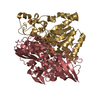
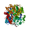
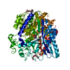
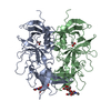
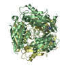
| Title | Structural insights into putative molybdenum cofactor biosynthesis protein C (MoaC2) from Mycobacterium tuberculosis H37Rv | ||||||
 Components Components | Molybdenum cofactor biosynthesis protein C 2 | ||||||
 Keywords Keywords | BIOSYNTHETIC PROTEIN / MoaC2 / Ferrodoxin-like fold / Moco biosynthesis | ||||||
| Function / homology |  Function and homology information Function and homology informationcyclic pyranopterin monophosphate synthase / cyclic pyranopterin monophosphate synthase activity / Mo-molybdopterin cofactor biosynthetic process Similarity search - Function | ||||||
| Biological species |   Mycobacterium tuberculosis (bacteria) Mycobacterium tuberculosis (bacteria) | ||||||
| Method |  X-RAY DIFFRACTION / SYNCHROTRON / MOLECULAR REPLACEMENT / Resolution: 2.202 Å X-RAY DIFFRACTION / SYNCHROTRON / MOLECULAR REPLACEMENT / Resolution: 2.202 Å | ||||||
 Authors Authors | Srivastava, V.K. / Srivastava, S. / Arora, A. / Pratap, J.V. | ||||||
 Citation Citation | Journal: Plos One / Year: 2013 Title: Structural Insights into Putative Molybdenum Cofactor Biosynthesis Protein C (MoaC2) from Mycobacterium tuberculosis H37Rv. Authors: Srivastava, V.K. / Srivastava, S. / Arora, A. / Pratap, J.V. | ||||||
| History |
|
- Structure visualization
| Structure viewer | Molecule: MolmilJmol/JSmol |
|---|
- Downloads & links
-Download
| PDBx/mmCIF format | 4fdf.cif.gz | 61.9 KB | Display | PDBx/mmCIF format |
|---|---|---|---|---|
| PDB format | pdb4fdf.ent.gz | 44.9 KB | Display | PDB format |
| PDBx/mmJSON format | 4fdf.json.gz | Tree view | PDBx/mmJSON format | |
| Others |  Other downloads Other downloads |
-Validation report
| Arichive directory | https://data.pdbj.org/pub/pdb/validation_reports/fd/4fdfftp://data.pdbj.org/pub/pdb/validation_reports/fd/4fdf | HTTPS FTP |
|---|
-Related structure data
| Related structure data |  1ekrS S: Starting model for refinement |
|---|---|
| Similar structure data |


-Links
 PDBj
PDBj- Assembly
| Deposited unit | 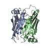
| ||||||||
|---|---|---|---|---|---|---|---|---|---|
| 1 | 
| ||||||||
| Unit cell |
|
-Components
| #1: Protein | Mass: 18693.270 Da / Num. of mol.: 2 Source method: isolated from a genetically manipulated source Source: (gene. exp.) Mycobacterium tuberculosis (bacteria) / Strain: H37Rv / Gene: M. tuberculosis, moaC2, MT0887, MTV043.57, Rv0864 / Plasmid: pET28b / Production host: #2: Water | ChemComp-HOH / |  Mass: 18.015 Da / Num. of mol.: 65 / Source method: isolated from a natural source / Formula: H2O Mass: 18.015 Da / Num. of mol.: 65 / Source method: isolated from a natural source / Formula: H2O |
|---|
-Experimental details
-Experiment
| Experiment | Method: X-RAY DIFFRACTION / Number of used crystals: 1 |
|---|
- Sample preparation
| Crystal | Density Matthews: 1.73 Å3/Da / Density % sol: 28.9 % |
|---|---|
| Crystal grow | Temperature: 295 K / Method: vapor diffusion, hanging drop / pH: 8 Details: 0.025M potassium sodium tartrate tetra hydrate solution, pH 8.0, VAPOR DIFFUSION, HANGING DROP, temperature 295K |
-Data collection
| Diffraction | Mean temperature: 100 K |
|---|---|
| Diffraction source | Source: SYNCHROTRON / Site: ESRF  / Beamline: BM14 / Wavelength: 0.976 Å / Beamline: BM14 / Wavelength: 0.976 Å |
| Detector | Type: MARRESEARCH / Detector: CCD / Date: Nov 18, 2011 |
| Radiation | Protocol: SINGLE WAVELENGTH / Monochromatic (M) / Laue (L): M / Scattering type: x-ray |
| Radiation wavelength | Wavelength: 0.976 Å / Relative weight: 1 |
| Reflection | Resolution: 2.2→27.71 Å / Num. obs: 13345 / % possible obs: 99.9 % / Observed criterion σ(F): 2 / Observed criterion σ(I): 1 |
| Reflection shell | Resolution: 2.2→2.28 Å / % possible all: 99.9 |
- Processing
| Software |
| ||||||||||||||||||||||||||||||||||||||||||||||||||||||||||||||||||||||||||||||||||||||||||||||||||||||||||||||||||||||||||||||||||||||||||||||||||||||||||||||||||||||||||
|---|---|---|---|---|---|---|---|---|---|---|---|---|---|---|---|---|---|---|---|---|---|---|---|---|---|---|---|---|---|---|---|---|---|---|---|---|---|---|---|---|---|---|---|---|---|---|---|---|---|---|---|---|---|---|---|---|---|---|---|---|---|---|---|---|---|---|---|---|---|---|---|---|---|---|---|---|---|---|---|---|---|---|---|---|---|---|---|---|---|---|---|---|---|---|---|---|---|---|---|---|---|---|---|---|---|---|---|---|---|---|---|---|---|---|---|---|---|---|---|---|---|---|---|---|---|---|---|---|---|---|---|---|---|---|---|---|---|---|---|---|---|---|---|---|---|---|---|---|---|---|---|---|---|---|---|---|---|---|---|---|---|---|---|---|---|---|---|---|---|---|---|
| Refinement | Method to determine structure: MOLECULAR REPLACEMENT Starting model: PDB ENTRY 1EKR Resolution: 2.202→27.71 Å / Cor.coef. Fo:Fc: 0.952 / Cor.coef. Fo:Fc free: 0.931 / SU B: 6.773 / SU ML: 0.173 / Cross valid method: THROUGHOUT / ESU R: 0.3 / ESU R Free: 0.222 / Stereochemistry target values: MAXIMUM LIKELIHOOD
| ||||||||||||||||||||||||||||||||||||||||||||||||||||||||||||||||||||||||||||||||||||||||||||||||||||||||||||||||||||||||||||||||||||||||||||||||||||||||||||||||||||||||||
| Solvent computation | Ion probe radii: 0.8 Å / Shrinkage radii: 0.8 Å / VDW probe radii: 1.2 Å / Solvent model: MASK | ||||||||||||||||||||||||||||||||||||||||||||||||||||||||||||||||||||||||||||||||||||||||||||||||||||||||||||||||||||||||||||||||||||||||||||||||||||||||||||||||||||||||||
| Displacement parameters | Biso mean: 45.449 Å2 | ||||||||||||||||||||||||||||||||||||||||||||||||||||||||||||||||||||||||||||||||||||||||||||||||||||||||||||||||||||||||||||||||||||||||||||||||||||||||||||||||||||||||||
| Refinement step | Cycle: LAST / Resolution: 2.202→27.71 Å
| ||||||||||||||||||||||||||||||||||||||||||||||||||||||||||||||||||||||||||||||||||||||||||||||||||||||||||||||||||||||||||||||||||||||||||||||||||||||||||||||||||||||||||
| Refine LS restraints |
| ||||||||||||||||||||||||||||||||||||||||||||||||||||||||||||||||||||||||||||||||||||||||||||||||||||||||||||||||||||||||||||||||||||||||||||||||||||||||||||||||||||||||||
| LS refinement shell | Resolution: 2.202→2.259 Å / Total num. of bins used: 20
|
