 Movie
Movie Controller

[English] 日本語
 Yorodumi
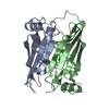
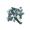
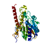
- PDB-6j0o: Crystal structure of CERT START domain in complex with compound SC1 -
+ Open data

- Basic information
| Entry | Database: PDB / ID: 6j0o | ||||||
|---|---|---|---|---|---|---|---|
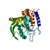
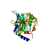
| Title | Crystal structure of CERT START domain in complex with compound SC1 | ||||||
 Components Components | LIPID-TRANSFER PROTEIN CERT | ||||||
 Keywords Keywords | LIPID TRANSPORT / CERT / PH / START / COMPLEX | ||||||
| Function / homology |  Function and homology information Function and homology informationintermembrane sphingolipid transfer / ceramide transfer activity / endoplasmic reticulum-trans-Golgi network membrane contact site / ER to Golgi ceramide transport / ceramide 1-phosphate transfer activity / ceramide 1-phosphate binding / sphingomyelin biosynthetic process / lipid transfer activity / intermembrane lipid transfer / ceramide metabolic process ...intermembrane sphingolipid transfer / ceramide transfer activity / endoplasmic reticulum-trans-Golgi network membrane contact site / ER to Golgi ceramide transport / ceramide 1-phosphate transfer activity / ceramide 1-phosphate binding / sphingomyelin biosynthetic process / lipid transfer activity / intermembrane lipid transfer / ceramide metabolic process / ceramide binding / Sphingolipid de novo biosynthesis / sphingolipid biosynthetic process / phosphatidylinositol-4-phosphate binding / kinase activity / immune response / endoplasmic reticulum membrane / Golgi apparatus / mitochondrion / nucleoplasm / cytosol Similarity search - Function | ||||||
| Biological species |  Homo sapiens (human) Homo sapiens (human) | ||||||
| Method |  X-RAY DIFFRACTION / MOLECULAR REPLACEMENT / Resolution: 1.8 Å X-RAY DIFFRACTION / MOLECULAR REPLACEMENT / Resolution: 1.8 Å | ||||||
 Authors Authors | Suzuki, M. / Nakao, N. / Ueno, M. / Sakai, S. / Egawa, D. / Hanzawa, H. / Kawasaki, S. / Kumagai, K. / Kobayashi, S. / Hanada, K. | ||||||
 Citation Citation | Journal: Commun Chem / Year: 2019 Title: Natural ligand-nonmimetic inhibitors of the lipid-transfer protein CERT Authors: Nakao, N. / Ueno, M. / Sakai, S. / Egawa, D. / Hanzawa, H. / Kawasaki, S. / Kumagai, K. / Suzuki, M. / Kobayashi, S. / Hanada, K. | ||||||
| History |
|
- Structure visualization
| Structure viewer | Molecule: MolmilJmol/JSmol |
|---|
- Downloads & links
-Download
| PDBx/mmCIF format | 6j0o.cif.gz | 67.1 KB | Display | PDBx/mmCIF format |
|---|---|---|---|---|
| PDB format | pdb6j0o.ent.gz | 46.8 KB | Display | PDB format |
| PDBx/mmJSON format | 6j0o.json.gz | Tree view | PDBx/mmJSON format | |
| Others |  Other downloads Other downloads |
-Validation report
| Arichive directory | https://data.pdbj.org/pub/pdb/validation_reports/j0/6j0oftp://data.pdbj.org/pub/pdb/validation_reports/j0/6j0o | HTTPS FTP |
|---|
-Related structure data
| Related structure data |  5zygC  5zyhC  5zyiC  5zyjC  5zykC  5zylC  5zymC 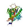 6iezC  6if0C  6j81C C: citing same article ( |
|---|---|
| Similar structure data |


-Links
 PDBj
PDBj
- Assembly
| Deposited unit | 
| ||||||||
|---|---|---|---|---|---|---|---|---|---|
| 1 |
| ||||||||
| Unit cell |
|
-Components
| #1: Protein | Mass: 27056.781 Da / Num. of mol.: 1 Source method: isolated from a genetically manipulated source Source: (gene. exp.) Homo sapiens (human) / Gene: CERT / Production host:  |
|---|---|
| #2: Chemical | ChemComp-XAF /   Mass: 437.487 Da / Num. of mol.: 1 / Source method: obtained synthetically / Formula: C23H20FN3O3S / Feature type: SUBJECT OF INVESTIGATION Mass: 437.487 Da / Num. of mol.: 1 / Source method: obtained synthetically / Formula: C23H20FN3O3S / Feature type: SUBJECT OF INVESTIGATION |
| #3: Chemical | ChemComp-UNX /   Num. of mol.: 1 / Source method: obtained synthetically Num. of mol.: 1 / Source method: obtained synthetically |
| #4: Water | ChemComp-HOH /  Mass: 18.015 Da / Num. of mol.: 208 / Source method: isolated from a natural source / Formula: H2O Mass: 18.015 Da / Num. of mol.: 208 / Source method: isolated from a natural source / Formula: H2O |
| Nonpolymer details | UNX 601 was modeled on an electron density peak clearly observed near methylpyridine moiety of XAF. ...UNX 601 was modeled on an electron density peak clearly observed near methylpyridine moiety of XAF. However distances between this peak and surrounding H-bond donor/acceptor are too short. Therefore it is assigned to unknown atom, not to water oxygen. |
| Sequence details | Authors state that these amino residues are originated from protease site after N-terminal affinity tag. |
-Experimental details
-Experiment
| Experiment | Method: X-RAY DIFFRACTION / Number of used crystals: 1 |
|---|
- Sample preparation
| Crystal | Density Matthews: 2.58 Å3/Da / Density % sol: 52.24 % |
|---|---|
| Crystal grow | Temperature: 293 K / Method: vapor diffusion, sitting drop / pH: 5.9 Details: 0.1M trisodium citrate/HCL buffer, pH5.9 containing 24% PEG3350 and 0.2% n-octyl-beta-D-glucoside |
-Data collection
| Diffraction | Mean temperature: 100 K / Serial crystal experiment: N | ||||||||||||||||||||||||||||||
|---|---|---|---|---|---|---|---|---|---|---|---|---|---|---|---|---|---|---|---|---|---|---|---|---|---|---|---|---|---|---|---|
| Diffraction source | Source: ROTATING ANODE / Type: RIGAKU MICROMAX-007 HF / Wavelength: 1.5418 Å | ||||||||||||||||||||||||||||||
| Detector | Type: RIGAKU RAXIS VII / Detector: IMAGE PLATE / Date: Apr 10, 2014 | ||||||||||||||||||||||||||||||
| Radiation | Monochromator: mirror / Protocol: SINGLE WAVELENGTH / Monochromatic (M) / Laue (L): M / Scattering type: x-ray | ||||||||||||||||||||||||||||||
| Radiation wavelength | Wavelength: 1.5418 Å / Relative weight: 1 | ||||||||||||||||||||||||||||||
| Reflection | Resolution: 1.8→30.13 Å / Num. obs: 27078 / % possible obs: 99.8 % / Redundancy: 4.5 % / CC1/2: 0.999 / Rmerge(I) obs: 0.047 / Rpim(I) all: 0.024 / Rrim(I) all: 0.053 / Net I/σ(I): 16.4 / Num. measured all: 122989 / Scaling rejects: 5 | ||||||||||||||||||||||||||||||
| Reflection shell | Diffraction-ID: 1
|
- Processing
| Software |
| |||||||||||||||||||||||||||||||||||||||||||||
|---|---|---|---|---|---|---|---|---|---|---|---|---|---|---|---|---|---|---|---|---|---|---|---|---|---|---|---|---|---|---|---|---|---|---|---|---|---|---|---|---|---|---|---|---|---|---|
| Refinement | Method to determine structure: MOLECULAR REPLACEMENT / Resolution: 1.8→25 Å / Cor.coef. Fo:Fc: 0.947 / Cor.coef. Fo:Fc free: 0.93 / SU B: 2.617 / SU ML: 0.083 / Cross valid method: THROUGHOUT / σ(F): 0 / ESU R: 0.127 / ESU R Free: 0.126 / Stereochemistry target values: MAXIMUM LIKELIHOOD / Details: U VALUES : REFINED INDIVIDUALLY
| |||||||||||||||||||||||||||||||||||||||||||||
| Solvent computation | Ion probe radii: 0.8 Å / Shrinkage radii: 0.8 Å / VDW probe radii: 1.2 Å / Solvent model: MASK | |||||||||||||||||||||||||||||||||||||||||||||
| Displacement parameters | Biso max: 78.37 Å2 / Biso mean: 26.48 Å2 / Biso min: 13.38 Å2
| |||||||||||||||||||||||||||||||||||||||||||||
| Refinement step | Cycle: final / Resolution: 1.8→25 Å
| |||||||||||||||||||||||||||||||||||||||||||||
| Refine LS restraints |
| |||||||||||||||||||||||||||||||||||||||||||||
| LS refinement shell | Resolution: 1.8→1.863 Å / Rfactor Rfree error: 0 / Total num. of bins used: 15
|
