 Movie
Movie Controller


+ Open data

- Basic information
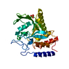
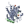
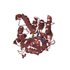
| Entry | Database: PDB / ID: 5ko9 | ||||||
|---|---|---|---|---|---|---|---|
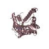
| Title | Crystal Structure of the SRAP Domain of Human HMCES Protein | ||||||
 Components Components | Embryonic stem cell-specific 5-hydroxymethylcytosine-binding protein | ||||||
 Keywords Keywords | DNA BINDING PROTEIN / SRAP Domain / DNA-binding / Structural Genomics / Structural Genomics Consortium / SGC | ||||||
| Function / homology |  Function and homology information Function and homology informationprotein-DNA covalent cross-linking activity / Lyases / DNA-(abasic site) binding / positive regulation of isotype switching / double-strand break repair via alternative nonhomologous end joining / protein-DNA covalent cross-linking repair / Hydrolases; Acting on peptide bonds (peptidases) / somatic hypermutation of immunoglobulin genes / interstrand cross-link repair / replication fork ...protein-DNA covalent cross-linking activity / Lyases / DNA-(abasic site) binding / positive regulation of isotype switching / double-strand break repair via alternative nonhomologous end joining / protein-DNA covalent cross-linking repair / Hydrolases; Acting on peptide bonds (peptidases) / somatic hypermutation of immunoglobulin genes / interstrand cross-link repair / replication fork / peptidase activity / single-stranded DNA binding / DNA damage response / proteolysis Similarity search - Function | ||||||
| Biological species |  Homo sapiens (human) Homo sapiens (human) | ||||||
| Method |  X-RAY DIFFRACTION / SYNCHROTRON / MOLECULAR REPLACEMENT / Resolution: 1.5 Å X-RAY DIFFRACTION / SYNCHROTRON / MOLECULAR REPLACEMENT / Resolution: 1.5 Å | ||||||
 Authors Authors | Halabelian, L. / Li, Y. / Bountra, C. / Edwards, A.M. / Arrowsmith, C.H. / Structural Genomics Consortium (SGC) | ||||||
 Citation Citation | Journal: Nat.Struct.Mol.Biol. / Year: 2019 Title: Structural basis of HMCES interactions with abasic DNA and multivalent substrate recognition. Authors: Halabelian, L. / Ravichandran, M. / Li, Y. / Zeng, H. / Rao, A. / Aravind, L. / Arrowsmith, C.H. | ||||||
| History |
|
- Structure visualization
| Structure viewer | Molecule: MolmilJmol/JSmol |
|---|
- Downloads & links
-Download
| PDBx/mmCIF format | 5ko9.cif.gz | 72.9 KB | Display | PDBx/mmCIF format |
|---|---|---|---|---|
| PDB format | pdb5ko9.ent.gz | 51.5 KB | Display | PDB format |
| PDBx/mmJSON format | 5ko9.json.gz | Tree view | PDBx/mmJSON format | |
| Others |  Other downloads Other downloads |
-Validation report
| Arichive directory | https://data.pdbj.org/pub/pdb/validation_reports/ko/5ko9ftp://data.pdbj.org/pub/pdb/validation_reports/ko/5ko9 | HTTPS FTP |
|---|
-Related structure data
| Related structure data |  6oe7C  6oeaC 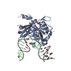 6oebC  1zn6S  2bdvS  2f20S  2icuS S: Starting model for refinement C: citing same article ( |
|---|---|
| Similar structure data |


-Links
 PDBj
PDBj- Assembly
| Deposited unit | 
| ||||||||
|---|---|---|---|---|---|---|---|---|---|
| 1 |
| ||||||||
| Unit cell |
|
-Components
| #1: Protein | Mass: 31802.816 Da / Num. of mol.: 1 Source method: isolated from a genetically manipulated source Source: (gene. exp.) Homo sapiens (human) / Gene: HMCES, C3orf37, DC12, SRAPD1 / Production host:  References: UniProt: Q96FZ2, Hydrolases; Acting on peptide bonds (peptidases) | ||
|---|---|---|---|
| #2: Chemical | ChemComp-UNX /   Num. of mol.: 44 / Source method: obtained synthetically Num. of mol.: 44 / Source method: obtained synthetically#3: Water | ChemComp-HOH / |  Mass: 18.015 Da / Num. of mol.: 186 / Source method: isolated from a natural source / Formula: H2O Mass: 18.015 Da / Num. of mol.: 186 / Source method: isolated from a natural source / Formula: H2O |
-Experimental details
-Experiment
| Experiment | Method: X-RAY DIFFRACTION / Number of used crystals: 1 |
|---|
- Sample preparation
| Crystal | Density Matthews: 2.44 Å3/Da / Density % sol: 44.89 % |
|---|---|
| Crystal grow | Temperature: 293 K / Method: vapor diffusion, sitting drop / pH: 6.5 / Details: 0.1M BTP, 2% Tacsimate, 20% PEG3350 |
-Data collection
| Diffraction | Mean temperature: 100 K | |||||||||||||||||||||
|---|---|---|---|---|---|---|---|---|---|---|---|---|---|---|---|---|---|---|---|---|---|---|
| Diffraction source | Source: SYNCHROTRON / Site: APS  / Beamline: 19-ID / Wavelength: 0.97926 Å / Beamline: 19-ID / Wavelength: 0.97926 Å | |||||||||||||||||||||
| Detector | Type: DECTRIS PILATUS3 6M / Detector: PIXEL / Date: Jun 15, 2016 | |||||||||||||||||||||
| Radiation | Protocol: SINGLE WAVELENGTH / Monochromatic (M) / Laue (L): M / Scattering type: x-ray | |||||||||||||||||||||
| Radiation wavelength | Wavelength: 0.97926 Å / Relative weight: 1 | |||||||||||||||||||||
| Reflection | Resolution: 1.5→48.37 Å / Num. obs: 43639 / % possible obs: 97 % / Redundancy: 3.8 % / CC1/2: 0.999 / Rmerge(I) obs: 0.045 / Net I/σ(I): 15.9 | |||||||||||||||||||||
| Reflection shell |
|
- Processing
| Software |
| |||||||||||||||||||||||||||||||||||||||||||||||||||||||||||||||||||||||||||
|---|---|---|---|---|---|---|---|---|---|---|---|---|---|---|---|---|---|---|---|---|---|---|---|---|---|---|---|---|---|---|---|---|---|---|---|---|---|---|---|---|---|---|---|---|---|---|---|---|---|---|---|---|---|---|---|---|---|---|---|---|---|---|---|---|---|---|---|---|---|---|---|---|---|---|---|---|
| Refinement | Method to determine structure: MOLECULAR REPLACEMENT Starting model: 2F20, 2BDV, 2ICU, 1ZN6 Resolution: 1.5→48.37 Å / Cor.coef. Fo:Fc: 0.964 / Cor.coef. Fo:Fc free: 0.954 / SU B: 1.549 / SU ML: 0.056 / Cross valid method: THROUGHOUT / σ(F): 0 / ESU R: 0.075 / ESU R Free: 0.076
| |||||||||||||||||||||||||||||||||||||||||||||||||||||||||||||||||||||||||||
| Solvent computation | Ion probe radii: 0.8 Å / Shrinkage radii: 0.8 Å / VDW probe radii: 1.2 Å | |||||||||||||||||||||||||||||||||||||||||||||||||||||||||||||||||||||||||||
| Displacement parameters | Biso max: 83.46 Å2 / Biso mean: 24.169 Å2 / Biso min: 12.43 Å2
| |||||||||||||||||||||||||||||||||||||||||||||||||||||||||||||||||||||||||||
| Refinement step | Cycle: final / Resolution: 1.5→48.37 Å
| |||||||||||||||||||||||||||||||||||||||||||||||||||||||||||||||||||||||||||
| Refine LS restraints |
| |||||||||||||||||||||||||||||||||||||||||||||||||||||||||||||||||||||||||||
| LS refinement shell | Resolution: 1.5→1.539 Å / Total num. of bins used: 20
|
