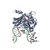
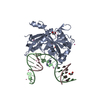
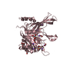
A: Embryonic stem cell-specific 5-hydroxymethylcytosine-binding protein C: DNA (5'-D(*GP*TP*CP*TP*GP*G)-3') B: DNA (5'-D(*CP*CP*AP*GP*AP*CP*GP*TP*(DRZ)P*GP*TP*T)-3') hetero molecules
A: Embryonic stem cell-specific 5-hydroxymethylcytosine-binding protein C: DNA (5'-D(*GP*TP*CP*TP*GP*G)-3') B: DNA (5'-D(*CP*CP*AP*GP*AP*CP*GP*TP*(DRZ)P*GP*TP*T)-3') hetero molecules
C: DNA (5'-D(*GP*TP*CP*TP*GP*G)-3') B: DNA (5'-D(*CP*CP*AP*GP*AP*CP*GP*TP*(DRZ)P*GP*TP*T)-3')
Embryonicstemcell-specific5-hydroxymethylcytosine-bindingprotein / ES cell-specific 5hmC-binding protein / Putative peptidase SRAPD1 / SRAP domain-containing protein 1
Mass: 31671.617 Da / Num. of mol.: 1 / Fragment: SRAP domain (UNP residues 2-270) Source method: isolated from a genetically manipulated source Source: (gene. exp.) Homo sapiens (human) / Gene: HMCES, C3orf37, DC12, SRAPD1 / Production host: Escherichia coli (E. coli) / References: UniProt: Q96FZ2
-
DNA chain , 2 types, 2 molecules CB
#2: DNA chain
DNA (5'-D(*GP*TP*CP*TP*GP*G)-3')
Mass: 1840.227 Da / Num. of mol.: 1 / Source method: obtained synthetically / Source: (synth.) synthetic construct (others)
#3: DNA chain
DNA (5'-D(*CP*CP*AP*GP*AP*CP*GP*TP*(DRZ)P*GP*TP*T)-3')
Mass: 3545.292 Da / Num. of mol.: 1 / Source method: obtained synthetically / Source: (synth.) synthetic construct (others)
Method to determine structure: FOURIER SYNTHESIS / Resolution: 2.2→48.72 Å / Cor.coef. Fo:Fc: 0.966 / Cor.coef. Fo:Fc free: 0.956 / SU B: 7.014 / SU ML: 0.159 / SU R Cruickshank DPI: 0.2117 / Cross valid method: THROUGHOUT / σ(F): 0 / ESU R: 0.212 / ESU R Free: 0.17 Details: HYDROGENS HAVE BEEN ADDED IN THE RIDING POSITIONS U VALUES : REFINED INDIVIDUALLY
Rfactor
Num. reflection
% reflection
Selection details
Rfree
0.2161
1036
4.9 %
RANDOM
Rwork
0.1942
-
-
-
obs
0.1954
19914
96.44 %
-
Solvent computation
Ion probe radii: 0.8 Å / Shrinkage radii: 0.8 Å / VDW probe radii: 1.2 Å
In the structure databanks used in Yorodumi, some data are registered as the other names, "COVID-19 virus" and "2019-nCoV". Here are the details of the virus and the list of structure data.
Jan 31, 2019. EMDB accession codes are about to change! (news from PDBe EMDB page)
EMDB accession codes are about to change! (news from PDBe EMDB page)
The allocation of 4 digits for EMDB accession codes will soon come to an end. Whilst these codes will remain in use, new EMDB accession codes will include an additional digit and will expand incrementally as the available range of codes is exhausted. The current 4-digit format prefixed with “EMD-” (i.e. EMD-XXXX) will advance to a 5-digit format (i.e. EMD-XXXXX), and so on. It is currently estimated that the 4-digit codes will be depleted around Spring 2019, at which point the 5-digit format will come into force.
The EM Navigator/Yorodumi systems omit the EMD- prefix.
Related info.:Q: What is EMD? / ID/Accession-code notation in Yorodumi/EM Navigator
Yorodumi is a browser for structure data from EMDB, PDB, SASBDB, etc.
This page is also the successor to EM Navigator detail page, and also detail information page/front-end page for Omokage search.
The word "yorodu" (or yorozu) is an old Japanese word meaning "ten thousand". "mi" (miru) is to see.
Related info.:EMDB / PDB / SASBDB / Comparison of 3 databanks / Yorodumi Search / Aug 31, 2016. New EM Navigator & Yorodumi / Yorodumi Papers / Jmol/JSmol / Function and homology information / Changes in new EM Navigator and Yorodumi
 Movie
Movie Controller


 Open data
 Basic information
 Components
 Keywords
 Function and homology information
 Homo sapiens (human)
 X-RAY DIFFRACTION /
  Authors
 Citation
 Structure visualization
 Downloads & links
 Other downloads


 PDBj
PDBj


 Assembly


 Num. of mol.: 4 / Source method: obtained synthetically
 Mass: 62.068 Da / Num. of mol.: 5 / Source method: obtained synthetically / Formula: C2H6O2
 Sample preparation
 / Beamline: 24-ID-C / Wavelength: 0.9789 Å
 Processing