 Movie
Movie Controller

[English] 日本語
 Yorodumi
- PDB-1xdk: Crystal Structure of the RARbeta/RXRalpha Ligand Binding Domain H... -
+ Open data

- Basic information
| Entry | Database: PDB / ID: 1xdk | ||||||
|---|---|---|---|---|---|---|---|
| Title | Crystal Structure of the RARbeta/RXRalpha Ligand Binding Domain Heterodimer in Complex with 9-cis Retinoic Acid and a Fragment of the TRAP220 Coactivator | ||||||
 Components Components |
| ||||||
 Keywords Keywords | HORMONE/GROWTH FACTOR RECEPTOR / Nuclear Receptor / Coactivator / Ligand / HORMONE-GROWTH FACTOR RECEPTOR COMPLEX | ||||||
| Function / homology |  Function and homology information Function and homology informationTranscriptional regulation of granulopoiesis / Carnitine shuttle / Transcriptional regulation of white adipocyte differentiation / Signaling by Retinoic Acid / NR1H2 & NR1H3 regulate gene expression to control bile acid homeostasis / NR1H3 & NR1H2 regulate gene expression linked to cholesterol transport and efflux / SUMOylation of intracellular receptors / Recycling of bile acids and salts / Synthesis of bile acids and bile salts / glandular epithelial cell development ...Transcriptional regulation of granulopoiesis / Carnitine shuttle / Transcriptional regulation of white adipocyte differentiation / Signaling by Retinoic Acid / NR1H2 & NR1H3 regulate gene expression to control bile acid homeostasis / NR1H3 & NR1H2 regulate gene expression linked to cholesterol transport and efflux / SUMOylation of intracellular receptors / Recycling of bile acids and salts / Synthesis of bile acids and bile salts / glandular epithelial cell development / visceral serous pericardium development / Nuclear Receptor transcription pathway / Synthesis of bile acids and bile salts via 7alpha-hydroxycholesterol / Synthesis of bile acids and bile salts via 27-hydroxycholesterol / embryonic eye morphogenesis / ventricular cardiac muscle cell differentiation / mesenchyme development / Endogenous sterols / positive regulation of translational initiation by iron / MLL4 and MLL3 complexes regulate expression of PPARG target genes in adipogenesis and hepatic steatosis / maternal placenta development / growth plate cartilage development / enucleate erythrocyte development / positive regulation of type II interferon-mediated signaling pathway / androgen biosynthetic process / positive regulation of G0 to G1 transition / angiogenesis involved in coronary vascular morphogenesis / regulation of RNA biosynthetic process / Regulation of lipid metabolism by PPARalpha / retinal pigment epithelium development / retinoic acid-responsive element binding / negative regulation of cartilage development / thyroid hormone receptor signaling pathway / Cytoprotection by HMOX1 / mammary gland branching involved in thelarche / nuclear protein quality control by the ubiquitin-proteasome system / secretory columnal luminar epithelial cell differentiation involved in prostate glandular acinus development / positive regulation of thyroid hormone receptor signaling pathway / core mediator complex / Estrogen-dependent gene expression / regulation of vitamin D receptor signaling pathway / embryonic digestive tract development / striatum development / ventricular trabecula myocardium morphogenesis / positive regulation of hepatocyte proliferation / positive regulation of keratinocyte differentiation / mediator complex / retinoic acid binding / thyroid hormone generation / nuclear retinoic acid receptor binding / camera-type eye development / outflow tract septum morphogenesis / embryonic heart tube development / cardiac muscle cell differentiation / cellular response to thyroid hormone stimulus / positive regulation of vitamin D receptor signaling pathway / negative regulation of chondrocyte differentiation / embryonic hindlimb morphogenesis / nuclear vitamin D receptor binding / positive regulation of programmed cell death / peroxisome proliferator activated receptor binding / lens development in camera-type eye / nuclear thyroid hormone receptor binding / heterocyclic compound binding / embryonic hemopoiesis / ureteric bud development / neural precursor cell proliferation / mammary gland epithelial cell proliferation / RNA polymerase II intronic transcription regulatory region sequence-specific DNA binding / regulation of myelination / regulation of branching involved in prostate gland morphogenesis / megakaryocyte development / cellular response to hepatocyte growth factor stimulus / DNA binding domain binding / cellular response to steroid hormone stimulus / positive regulation of intracellular estrogen receptor signaling pathway / epithelial cell proliferation involved in mammary gland duct elongation / ventricular cardiac muscle tissue morphogenesis / negative regulation of neuron differentiation / histone acetyltransferase binding / LBD domain binding / mammary gland branching involved in pregnancy / positive regulation of lipoprotein transport / nuclear steroid receptor activity / nuclear receptor-mediated steroid hormone signaling pathway / animal organ regeneration / negative regulation of keratinocyte proliferation / monocyte differentiation / general transcription initiation factor binding / response to retinoic acid / cellular response to low-density lipoprotein particle stimulus / embryonic placenta development / positive regulation of bone mineralization / cardiac muscle cell proliferation / positive regulation of transcription initiation by RNA polymerase II / nuclear retinoid X receptor binding / fat cell differentiation / ubiquitin ligase complex / RNA polymerase II preinitiation complex assembly / retinoic acid receptor signaling pathway Similarity search - Function | ||||||
| Biological species |  | ||||||
| Method |  X-RAY DIFFRACTION / SYNCHROTRON / MOLECULAR REPLACEMENT / Resolution: 2.9 Å X-RAY DIFFRACTION / SYNCHROTRON / MOLECULAR REPLACEMENT / Resolution: 2.9 Å | ||||||
 Authors Authors | Pogenberg, V. / Guichou, J.F. / Vivat-Hannah, V. / Kammerer, S. / Perez, E. / Germain, P. / De Lera, A.R. / Gronemeyer, H. / Royer, C.A. / Bourguet, W. | ||||||
 Citation Citation | Journal: J.Biol.Chem. / Year: 2005 Title: CHARACTERIZATION OF THE INTERACTION BETWEEN RAR/RXR HETERODIMERS AND TRANSCRIPTIONAL COACTIVATORS THROUGH STRUCTURAL AND FLUORESCENCE ANISOTROPY STUDIES Authors: Pogenberg, V. / Guichou, J.F. / Vivat-Hannah, V. / Kammerer, S. / Perez, E. / Germain, P. / De Lera, A.R. / Gronemeyer, H. / Royer, C.A. / Bourguet, W. | ||||||
| History |
|
- Structure visualization
| Structure viewer | Molecule: MolmilJmol/JSmol |
|---|
- Downloads & links
-Download
| PDBx/mmCIF format | 1xdk.cif.gz | 203.2 KB | Display | PDBx/mmCIF format |
|---|---|---|---|---|
| PDB format | pdb1xdk.ent.gz | 161.3 KB | Display | PDB format |
| PDBx/mmJSON format | 1xdk.json.gz | Tree view | PDBx/mmJSON format | |
| Others |  Other downloads Other downloads |
-Validation report
| Arichive directory | https://data.pdbj.org/pub/pdb/validation_reports/xd/1xdkftp://data.pdbj.org/pub/pdb/validation_reports/xd/1xdk | HTTPS FTP |
|---|
-Related structure data
| Related structure data |  1dkfS S: Starting model for refinement |
|---|---|
| Similar structure data |


-Links
 PDBj
PDBj


- Assembly
| Deposited unit | 
| ||||||||
|---|---|---|---|---|---|---|---|---|---|
| 1 | 
| ||||||||
| 2 | 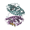
| ||||||||
| 3 |
| ||||||||
| Unit cell |
|
-Components
| #1: Protein | Mass: 26579.727 Da / Num. of mol.: 2 / Fragment: Ligand-Binding Domain Source method: isolated from a genetically manipulated source Source: (gene. exp.)  #2: Protein | Mass: 33978.113 Da / Num. of mol.: 2 / Fragment: Ligand-Binding Domain Source method: isolated from a genetically manipulated source Source: (gene. exp.) #3: Protein/peptide | Mass: 1609.910 Da / Num. of mol.: 4 / Fragment: Nuclear Receptor Box 2 / Source method: obtained synthetically Details: The peptide was chemically synthesized. The sequence of the peptide is naturally found in Mus musculus (mouse). References: UniProt: Q8BX19, UniProt: Q925J9*PLUS #4: Chemical | ChemComp-9CR / (   Mass: 300.435 Da / Num. of mol.: 4 / Source method: obtained synthetically / Formula: C20H28O2 / Comment: anticancer, antineoplastic*YM Mass: 300.435 Da / Num. of mol.: 4 / Source method: obtained synthetically / Formula: C20H28O2 / Comment: anticancer, antineoplastic*YM#5: Water | ChemComp-HOH / |  Mass: 18.015 Da / Num. of mol.: 12 / Source method: isolated from a natural source / Formula: H2O Mass: 18.015 Da / Num. of mol.: 12 / Source method: isolated from a natural source / Formula: H2O |
|---|
-Experimental details
-Experiment
| Experiment | Method: X-RAY DIFFRACTION / Number of used crystals: 1 |
|---|
- Sample preparation
| Crystal | Density Matthews: 3.6 Å3/Da / Density % sol: 65.5 % |
|---|---|
| Crystal grow | Temperature: 291 K / pH: 7.5 Details: PEG 3350, Sodium Formate, pH 7.5, VAPOR DIFFUSION, HANGING DROP, temperature 291K, pH 7.50 |
-Data collection
| Diffraction | Mean temperature: 100 K |
|---|---|
| Diffraction source | Source: SYNCHROTRON / Site: ESRF  / Beamline: BM30A / Wavelength: 0.9797 / Beamline: BM30A / Wavelength: 0.9797 |
| Detector | Type: MARRESEARCH / Detector: CCD / Date: Nov 19, 2003 |
| Radiation | Monochromator: SILICON / Protocol: SINGLE WAVELENGTH / Monochromatic (M) / Laue (L): M / Scattering type: x-ray |
| Radiation wavelength | Wavelength: 0.9797 Å / Relative weight: 1 |
| Reflection | Resolution: 2.9→36.21 Å / Num. obs: 42989 / % possible obs: 99.7 % / Redundancy: 7.4 % / Biso Wilson estimate: 92 Å2 / Rsym value: 0.083 / Net I/σ(I): 5.2 |
| Reflection shell | Resolution: 2.9→3.06 Å / Redundancy: 7.5 % / Mean I/σ(I) obs: 2 / Rsym value: 0.38 / % possible all: 99.6 |
- Processing
| Software |
| ||||||||||||||||||||||||||||||||||||||||||||||||||||||||||||
|---|---|---|---|---|---|---|---|---|---|---|---|---|---|---|---|---|---|---|---|---|---|---|---|---|---|---|---|---|---|---|---|---|---|---|---|---|---|---|---|---|---|---|---|---|---|---|---|---|---|---|---|---|---|---|---|---|---|---|---|---|---|
| Refinement | Method to determine structure: MOLECULAR REPLACEMENT Starting model: TRUNCATED VERSION OF PDB ENTRY 1DKF Resolution: 2.9→30 Å / Isotropic thermal model: RESTRAINED / Cross valid method: THROUGHOUT / σ(F): 0 / Stereochemistry target values: ENGH & HUBER
| ||||||||||||||||||||||||||||||||||||||||||||||||||||||||||||
| Solvent computation | Bsol: 53.7918 Å2 / ksol: 0.345913 e/Å3 | ||||||||||||||||||||||||||||||||||||||||||||||||||||||||||||
| Displacement parameters | Biso mean: 73.3 Å2
| ||||||||||||||||||||||||||||||||||||||||||||||||||||||||||||
| Refine analyze | Luzzati coordinate error free: 0.56 Å / Luzzati sigma a free: 0.8 Å | ||||||||||||||||||||||||||||||||||||||||||||||||||||||||||||
| Refinement step | Cycle: LAST / Resolution: 2.9→30 Å
| ||||||||||||||||||||||||||||||||||||||||||||||||||||||||||||
| Refine LS restraints |
| ||||||||||||||||||||||||||||||||||||||||||||||||||||||||||||
| LS refinement shell | Resolution: 2.9→3.08 Å / Rfactor Rfree error: 0.028 / Total num. of bins used: 6
| ||||||||||||||||||||||||||||||||||||||||||||||||||||||||||||
| Xplor file | Serial no: 1 / Param file: PROTEIN_REP.PARAM / Topol file: PROTEIN.TOP |
