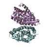
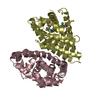
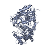
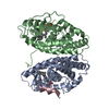
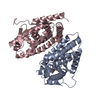
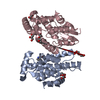
Entry Database : PDB / ID : 1uhlTitle Crystal structure of the LXRalfa-RXRbeta LBD heterodimer 10-mer peptide from Nuclear receptor coactivator 2 Oxysterols receptor LXR-alpha Retinoic acid receptor RXR-beta Keywords / Function / homology Function Domain/homology Component
/ / / / / / / / / / / / / / / / / / / / / / / / / / / / / / / / / / / / / / / / / / / / / / / / / / / / / / / / / / / / / / / / / / / / / / / / / / / / / / / / / / / / / / / / / / / / / / / / / / / / / / / / / / / / / / / / / / / / / / / / / / / / / / / / / / / / / / / / / / / / / / / / / / / / / / / / / / / / / / / / / / / / / / / Biological species Homo sapiens (human)Method / / / Resolution : 2.9 Å Authors Svensson, S. / Ostberg, T. / Jacobsson, M. / Norstrom, C. / Stefansson, K. / Hallen, D. / Johansson, I.C. / Zachrisson, K. / Ogg, D. / Jendeberg, L. Journal : Embo J. / Year : 2003Title : Crystal structure of the heterodimeric complex of LXRalpha and RXRbeta ligand-binding domains in a fully agonistic conformationAuthors : Svensson, S. / Ostberg, T. / Jacobsson, M. / Norstrom, C. / Stefansson, K. / Hallen, D. / Johansson, I.C. / Zachrisson, K. / Ogg, D. / Jendeberg, L. History Deposition Jul 3, 2003 Deposition site / Processing site Revision 1.0 Jun 1, 2004 Provider / Type Revision 1.1 Apr 27, 2008 Group Revision 1.2 Jul 13, 2011 Group Revision 1.3 May 23, 2018 Group / Category / Item Revision 1.4 Oct 25, 2023 Group Data collection / Database references ... Data collection / Database references / Derived calculations / Refinement description Category chem_comp_atom / chem_comp_bond ... chem_comp_atom / chem_comp_bond / database_2 / pdbx_initial_refinement_model / struct_ref_seq_dif / struct_site Item _database_2.pdbx_DOI / _database_2.pdbx_database_accession ... _database_2.pdbx_DOI / _database_2.pdbx_database_accession / _struct_ref_seq_dif.details / _struct_site.pdbx_auth_asym_id / _struct_site.pdbx_auth_comp_id / _struct_site.pdbx_auth_seq_id
Show all Show less
 Movie
Movie Controller


 Open data
 Basic information
 Components
 Keywords
 Function and homology information
 Homo sapiens (human)
 X-RAY DIFFRACTION /
  Authors
 Citation
 Structure visualization
 Downloads & links
 Other downloads
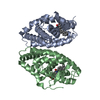
 PDBj
PDBj


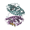
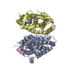
 Assembly

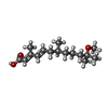

 Mass: 268.392 Da / Num. of mol.: 1 / Source method: obtained synthetically / Formula: C16H28O3
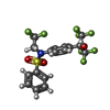
 Mass: 481.333 Da / Num. of mol.: 1 / Source method: obtained synthetically / Formula: C17H12F9NO3S
 Sample preparation
 / Beamline: I711 / Wavelength: 1.11 Å
 Processing