[English] 日本語
 Yorodumi
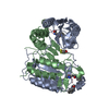
- PDB-1qdu: CRYSTAL STRUCTURE OF THE COMPLEX OF CASPASE-8 WITH THE TRIPEPTIDE... -
+ Open data
- Basic information
| Entry | Database: PDB / ID: 1qdu | ||||||
|---|---|---|---|---|---|---|---|
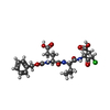
| Title | CRYSTAL STRUCTURE OF THE COMPLEX OF CASPASE-8 WITH THE TRIPEPTIDE KETONE INHIBITOR ZEVD-DCBMK | ||||||
 Components Components |
| ||||||
 Keywords Keywords | HYDROLASE/HYDROLASE INHIBITOR / APOPTOSIS / CYSTEINE PROTEASE / HYDROLASE-HYDROLASE INHIBITOR COMPLEX | ||||||
| Function / homology |  Function and homology information Function and homology informationcaspase-8 / syncytiotrophoblast cell differentiation involved in labyrinthine layer development / death effector domain binding / FasL/ CD95L signaling / TRAIL signaling / CD95 death-inducing signaling complex / Apoptotic execution phase / Activation, myristolyation of BID and translocation to mitochondria / ripoptosome / Defective RIPK1-mediated regulated necrosis ...caspase-8 / syncytiotrophoblast cell differentiation involved in labyrinthine layer development / death effector domain binding / FasL/ CD95L signaling / TRAIL signaling / CD95 death-inducing signaling complex / Apoptotic execution phase / Activation, myristolyation of BID and translocation to mitochondria / ripoptosome / Defective RIPK1-mediated regulated necrosis / Microbial modulation of RIPK1-mediated regulated necrosis / TRAIL-activated apoptotic signaling pathway / TRIF-mediated programmed cell death / Regulation by c-FLIP / CASP8 activity is inhibited / Dimerization of procaspase-8 / TLR3-mediated TICAM1-dependent programmed cell death / self proteolysis / Caspase activation via Death Receptors in the presence of ligand / positive regulation of macrophage differentiation / response to cobalt ion / NF-kB activation through FADD/RIP-1 pathway mediated by caspase-8 and -10 / death-inducing signaling complex / CLEC7A/inflammasome pathway / negative regulation of necroptotic process / regulation of tumor necrosis factor-mediated signaling pathway / tumor necrosis factor receptor binding / death receptor binding / natural killer cell activation / TNFR1-induced proapoptotic signaling / RIPK1-mediated regulated necrosis / execution phase of apoptosis / response to anesthetic / regulation of innate immune response / Apoptotic cleavage of cellular proteins / pyroptotic inflammatory response / response to tumor necrosis factor / B cell activation / macrophage differentiation / positive regulation of proteolysis / extrinsic apoptotic signaling pathway via death domain receptors / Caspase-mediated cleavage of cytoskeletal proteins / positive regulation of execution phase of apoptosis / extrinsic apoptotic signaling pathway / negative regulation of canonical NF-kappaB signal transduction / cysteine-type peptidase activity / regulation of cytokine production / : / T cell activation / protein maturation / positive regulation of interleukin-1 beta production / Regulation of NF-kappa B signaling / apoptotic signaling pathway / Regulation of TNFR1 signaling / cellular response to mechanical stimulus / NOD1/2 Signaling Pathway / protein processing / Regulation of necroptotic cell death / response to estradiol / peptidase activity / positive regulation of neuron apoptotic process / lamellipodium / heart development / cell body / scaffold protein binding / response to ethanol / angiogenesis / response to lipopolysaccharide / mitochondrial outer membrane / cytoskeleton / positive regulation of canonical NF-kappaB signal transduction / positive regulation of cell migration / positive regulation of apoptotic process / cysteine-type endopeptidase activity / apoptotic process / ubiquitin protein ligase binding / protein-containing complex binding / protein-containing complex / mitochondrion / proteolysis / identical protein binding / nucleus / cytosol / cytoplasm Similarity search - Function | ||||||
| Biological species |  Homo sapiens (human) Homo sapiens (human) | ||||||
| Method |  X-RAY DIFFRACTION / X-RAY DIFFRACTION /  SYNCHROTRON / SYNCHROTRON /  MOLECULAR REPLACEMENT / Resolution: 2.8 Å MOLECULAR REPLACEMENT / Resolution: 2.8 Å | ||||||
 Authors Authors | Blanchard, H. / Grutter, M.G. | ||||||
 Citation Citation |  Journal: Structure Fold.Des. / Year: 1999 Journal: Structure Fold.Des. / Year: 1999Title: The three-dimensional structure of caspase-8: an initiator enzyme in apoptosis. Authors: Blanchard, H. / Kodandapani, L. / Mittl, P.R. / Marco, S.D. / Krebs, J.F. / Wu, J.C. / Tomaselli, K.J. / Grutter, M.G. | ||||||
| History |
|
- Structure visualization
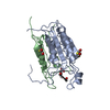
| Structure viewer | Molecule:  Molmil Molmil Jmol/JSmol Jmol/JSmol |
|---|
- Downloads & links
- Download
| PDBx/mmCIF format |  1qdu.cif.gz 1qdu.cif.gz | 272.2 KB | Display |  PDBx/mmCIF format PDBx/mmCIF format |
|---|---|---|---|---|
| PDB format |  pdb1qdu.ent.gz pdb1qdu.ent.gz | 222.6 KB | Display |  PDB format PDB format |
| PDBx/mmJSON format |  1qdu.json.gz 1qdu.json.gz | Tree view |  PDBx/mmJSON format PDBx/mmJSON format | |
| Others |  Other downloads Other downloads |
-Validation report
| Arichive directory |  https://data.pdbj.org/pub/pdb/validation_reports/qd/1qdu https://data.pdbj.org/pub/pdb/validation_reports/qd/1qdu ftp://data.pdbj.org/pub/pdb/validation_reports/qd/1qdu ftp://data.pdbj.org/pub/pdb/validation_reports/qd/1qdu | HTTPS FTP |
|---|
-Related structure data
| Similar structure data |
|---|
- Links
Links
- Assembly
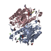
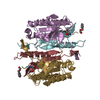
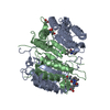
| Deposited unit | 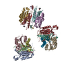
| ||||||||
|---|---|---|---|---|---|---|---|---|---|
| 1 | 
| ||||||||
| 2 | 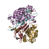
| ||||||||
| 3 | 
| ||||||||
| Unit cell |
|
- Components
| #1: Protein | Mass: 17416.957 Da / Num. of mol.: 6 Source method: isolated from a genetically manipulated source Source: (gene. exp.)  Homo sapiens (human) / Plasmid: PET21B / Production host: Homo sapiens (human) / Plasmid: PET21B / Production host:  References: UniProt: Q14790, Hydrolases; Acting on peptide bonds (peptidases); Cysteine endopeptidases #2: Protein | Mass: 10186.642 Da / Num. of mol.: 6 Source method: isolated from a genetically manipulated source Source: (gene. exp.)  Homo sapiens (human) / Plasmid: PET21B / Production host: Homo sapiens (human) / Plasmid: PET21B / Production host:  References: UniProt: Q14790, Hydrolases; Acting on peptide bonds (peptidases); Cysteine endopeptidases #3: Protein/peptide | #4: Water | ChemComp-HOH / | Compound details | THE UNBOUND INHIBITOR (CHAINS T,U,V,W,X,Y) IS CARBOBENZOXY-GLU-VAL-ASP-DICHLOROMETHYLKETONE. UPON ...THE UNBOUND INHIBITOR (CHAINS T,U,V,W,X,Y) IS CARBOBENZO | Has protein modification | Y | Sequence details | AMINO ACID RESIDUES ARE NUMBERED TO FACILITATE COMPARISON WITH CASPASE-1 (INTERLEUKIN 1-BETA ...AMINO ACID RESIDUES ARE NUMBERED TO FACILITATE | |
|---|
-Experimental details
-Experiment
| Experiment | Method:  X-RAY DIFFRACTION / Number of used crystals: 1 X-RAY DIFFRACTION / Number of used crystals: 1 |
|---|
- Sample preparation
| Crystal | Density Matthews: 2.86 Å3/Da / Density % sol: 57.02 % | |||||||||||||||||||||||||
|---|---|---|---|---|---|---|---|---|---|---|---|---|---|---|---|---|---|---|---|---|---|---|---|---|---|---|
| Crystal grow | Temperature: 277 K / Method: vapor diffusion, hanging drop / pH: 6.3 Details: 300mM ammonium phosphate, 27% (w/v) isopropanol, 100mM sodium phosphate, pH 6.3, VAPOR DIFFUSION, HANGING DROP, temperature 277K | |||||||||||||||||||||||||
| Crystal grow | *PLUS Temperature: 4 ℃ / Method: vapor diffusionDetails: drop consists of equal volume of protein and reservoir solutions | |||||||||||||||||||||||||
| Components of the solutions | *PLUS
|
-Data collection
| Diffraction | Mean temperature: 100 K |
|---|---|
| Diffraction source | Source:  SYNCHROTRON / Site: SYNCHROTRON / Site:  ESRF ESRF  / Beamline: BM1A / Wavelength: 0.873 / Beamline: BM1A / Wavelength: 0.873 |
| Detector | Type: MARRESEARCH / Detector: IMAGE PLATE / Date: Oct 2, 1997 |
| Radiation | Protocol: SINGLE WAVELENGTH / Monochromatic (M) / Laue (L): M / Scattering type: x-ray |
| Radiation wavelength | Wavelength: 0.873 Å / Relative weight: 1 |
| Reflection | Resolution: 2.8→100 Å / Num. all: 44902 / Num. obs: 44902 / % possible obs: 96.3 % / Observed criterion σ(I): 2 / Redundancy: 3.4 % / Rmerge(I) obs: 0.084 / Net I/σ(I): 17.1 |
| Reflection shell | Resolution: 2.8→2.85 Å / Redundancy: 3.1 % / Rmerge(I) obs: 0.469 / Mean I/σ(I) obs: 2.1 / Num. unique all: 2041 / % possible all: 84.7 |
| Reflection shell | *PLUS % possible obs: 84.7 % / Num. unique obs: 2041 |
- Processing
| Software |
| |||||||||||||||||||||||||
|---|---|---|---|---|---|---|---|---|---|---|---|---|---|---|---|---|---|---|---|---|---|---|---|---|---|---|
| Refinement | Method to determine structure:  MOLECULAR REPLACEMENT MOLECULAR REPLACEMENTStarting model: HOMOLOGY MODEL Resolution: 2.8→100 Å / Cross valid method: THROUGHOUT / σ(F): 0 / Stereochemistry target values: Engh & Huber
| |||||||||||||||||||||||||
| Refinement step | Cycle: LAST / Resolution: 2.8→100 Å
| |||||||||||||||||||||||||
| Refine LS restraints |
| |||||||||||||||||||||||||
| Software | *PLUS Name:  X-PLOR / Version: 3.851 / Classification: refinement X-PLOR / Version: 3.851 / Classification: refinement | |||||||||||||||||||||||||
| Refine LS restraints | *PLUS
|
 Movie
Movie Controller





 PDBj
PDBj


