caspase-6 / Breakdown of the nuclear lamina / regulation of programmed cell death / cellular response to staurosporine / positive regulation of necroptotic process / Apoptotic cleavage of cellular proteins / TP53 Regulates Transcription of Caspase Activators and Caspases / hepatocyte apoptotic process / protein autoprocessing / intrinsic apoptotic signaling pathway by p53 class mediator ...caspase-6 / Breakdown of the nuclear lamina / regulation of programmed cell death / cellular response to staurosporine / positive regulation of necroptotic process / Apoptotic cleavage of cellular proteins / TP53 Regulates Transcription of Caspase Activators and Caspases / hepatocyte apoptotic process / protein autoprocessing / intrinsic apoptotic signaling pathway by p53 class mediator / pyroptotic inflammatory response / Caspase-mediated cleavage of cytoskeletal proteins / epithelial cell differentiation / cysteine-type peptidase activity / activation of innate immune response / positive regulation of neuron apoptotic process / positive regulation of apoptotic process / cysteine-type endopeptidase activity / apoptotic process / proteolysis / nucleoplasm / identical protein binding / nucleus / cytoplasm / cytosol Similarity search - Function
Rossmann fold - #1460 / Peptidase C14 family / Peptidase family C14A, His active site / Caspase family histidine active site. / Peptidase C14, caspase non-catalytic subunit p10 / Peptidase family C14A, cysteine active site / Caspase family cysteine active site. / Caspase family p10 domain profile. / Peptidase C14A, caspase catalytic domain / Caspase, interleukin-1 beta converting enzyme (ICE) homologues ...Rossmann fold - #1460 / Peptidase C14 family / Peptidase family C14A, His active site / Caspase family histidine active site. / Peptidase C14, caspase non-catalytic subunit p10 / Peptidase family C14A, cysteine active site / Caspase family cysteine active site. / Caspase family p10 domain profile. / Peptidase C14A, caspase catalytic domain / Caspase, interleukin-1 beta converting enzyme (ICE) homologues / Peptidase C14, p20 domain / Caspase family p20 domain profile. / : / Caspase domain / Caspase-like domain superfamily / Rossmann fold / 3-Layer(aba) Sandwich / Alpha Beta Similarity search - Domain/homology
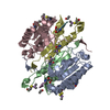
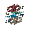
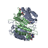
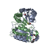
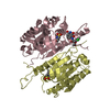
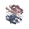
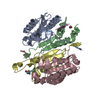
N-acetyl-L-valyl-L-alpha-glutamyl-N-[(2S)-1-carboxy-3-oxopropan-2-yl]-L-isoleucinamide / ACETATE ION / CACODYLATE ION / Caspase-6 Similarity search - Component
Biological species
Homo sapiens (human)
Method
X-RAY DIFFRACTION / SYNCHROTRON / SAD / Resolution: 1.625 Å
In the structure databanks used in Yorodumi, some data are registered as the other names, "COVID-19 virus" and "2019-nCoV". Here are the details of the virus and the list of structure data.
Jan 31, 2019. EMDB accession codes are about to change! (news from PDBe EMDB page)
EMDB accession codes are about to change! (news from PDBe EMDB page)
The allocation of 4 digits for EMDB accession codes will soon come to an end. Whilst these codes will remain in use, new EMDB accession codes will include an additional digit and will expand incrementally as the available range of codes is exhausted. The current 4-digit format prefixed with “EMD-” (i.e. EMD-XXXX) will advance to a 5-digit format (i.e. EMD-XXXXX), and so on. It is currently estimated that the 4-digit codes will be depleted around Spring 2019, at which point the 5-digit format will come into force.
The EM Navigator/Yorodumi systems omit the EMD- prefix.
Related info.:Q: What is EMD? / ID/Accession-code notation in Yorodumi/EM Navigator
Yorodumi is a browser for structure data from EMDB, PDB, SASBDB, etc.
This page is also the successor to EM Navigator detail page, and also detail information page/front-end page for Omokage search.
The word "yorodu" (or yorozu) is an old Japanese word meaning "ten thousand". "mi" (miru) is to see.
Related info.:EMDB / PDB / SASBDB / Comparison of 3 databanks / Yorodumi Search / Aug 31, 2016. New EM Navigator & Yorodumi / Yorodumi Papers / Jmol/JSmol / Function and homology information / Changes in new EM Navigator and Yorodumi
 Movie
Movie Controller

 Yorodumi
 Open data
 Basic information
 Components
 Keywords
 Function and homology information
 Homo sapiens (human)
 X-RAY DIFFRACTION /
  Authors
 Citation
 Structure visualization
 Downloads & links
 Other downloads
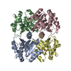


 PDBj
PDBj


 Assembly
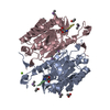

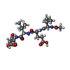
 Type: Polypeptide / Class: Inhibitor / Mass: 484.543 Da / Num. of mol.: 2 / Source method: obtained synthetically / Details: Chemically synthesized


 Mass: 136.989 Da / Num. of mol.: 8 / Source method: obtained synthetically / Formula: C2H6AsO2
 Mass: 59.044 Da / Num. of mol.: 1 / Source method: obtained synthetically / Formula: C2H3O2
 Mass: 24.305 Da / Num. of mol.: 1 / Source method: obtained synthetically / Formula: Mg
 Sample preparation
 / Beamline: BL-6A / Wavelength: 1 Å
 Processing