 Movie
Movie Controller

[English] 日本語
 Yorodumi
- PDB-1t06: 1.9 A Crystal Structure of a Protein of Unknown Function from Bac... -
+ Open data

- Basic information
| Entry | Database: PDB / ID: 1t06 | ||||||
|---|---|---|---|---|---|---|---|
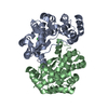
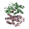
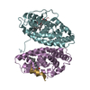
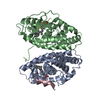
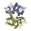
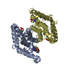
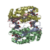
| Title | 1.9 A Crystal Structure of a Protein of Unknown Function from Bacillus cereus ATCC 14579 | ||||||
 Components Components | hypothetical protein | ||||||
 Keywords Keywords | STRUCTURAL GENOMICS / UNKNOWN FUNCTION / hypothetical protein / Bacillus cereus / PSI / Protein Structure Initiative / Midwest Center for Structural Genomics / MCSG | ||||||
| Function / homology | DNA alkylation repair enzyme / DNA alkylation repair enzyme / Armadillo-type fold / DNA alkylation repair protein Function and homology information Function and homology information | ||||||
| Biological species |  | ||||||
| Method |  X-RAY DIFFRACTION / SYNCHROTRON / SAD / Resolution: 1.9 Å X-RAY DIFFRACTION / SYNCHROTRON / SAD / Resolution: 1.9 Å | ||||||
 Authors Authors | Zhang, R. / Wu, R. / Moy, S. / Joachimiak, A. / Midwest Center for Structural Genomics (MCSG) | ||||||
 Citation Citation | Journal: To be Published Title: 1.9A crystal structure of a hypothetical protein from Bacillus cereus ATCC 14579 Authors: Zhang, R. / Wu, R. / Moy, S. / Joachimiak, A. | ||||||
| History |
|
- Structure visualization
| Structure viewer | Molecule: MolmilJmol/JSmol |
|---|
- Downloads & links
-Download
| PDBx/mmCIF format | 1t06.cif.gz | 109.5 KB | Display | PDBx/mmCIF format |
|---|---|---|---|---|
| PDB format | pdb1t06.ent.gz | 84.7 KB | Display | PDB format |
| PDBx/mmJSON format | 1t06.json.gz | Tree view | PDBx/mmJSON format | |
| Others |  Other downloads Other downloads |
-Validation report
| Arichive directory | https://data.pdbj.org/pub/pdb/validation_reports/t0/1t06ftp://data.pdbj.org/pub/pdb/validation_reports/t0/1t06 | HTTPS FTP |
|---|
-Related structure data
| Similar structure data | |
|---|---|
| Other databases |


-Links
 PDBj
PDBj- Assembly
| Deposited unit | 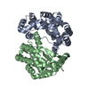
| ||||||||
|---|---|---|---|---|---|---|---|---|---|
| 1 |
| ||||||||
| Unit cell |
| ||||||||
| Details | This protein may existed in dimmer, MolA and MolB present the dimmer in the asymmetric unit. |
-Components
| #1: Protein | Mass: 26512.344 Da / Num. of mol.: 2 Source method: isolated from a genetically manipulated source Source: (gene. exp.) #2: Water | ChemComp-HOH / |  Mass: 18.015 Da / Num. of mol.: 405 / Source method: isolated from a natural source / Formula: H2O Mass: 18.015 Da / Num. of mol.: 405 / Source method: isolated from a natural source / Formula: H2O |
|---|
-Experimental details
-Experiment
| Experiment | Method: X-RAY DIFFRACTION / Number of used crystals: 1 |
|---|
- Sample preparation
| Crystal | Density Matthews: 2.35 Å3/Da / Density % sol: 44.54 % |
|---|---|
| Crystal grow | Temperature: 298 K / Method: vapor diffusion, sitting drop / pH: 5.5 Details: 1.2M NH4SO4, 20% PEG 4000, pH 5.5, VAPOR DIFFUSION, SITTING DROP, temperature 298K |
-Data collection
| Diffraction | Mean temperature: 100 K |
|---|---|
| Diffraction source | Source: SYNCHROTRON / Site: APS  / Beamline: 19-ID / Wavelength: 0.9795 Å / Beamline: 19-ID / Wavelength: 0.9795 Å |
| Detector | Type: SBC-2 / Detector: CCD / Date: Mar 30, 2004 / Details: mirrors |
| Radiation | Monochromator: Si 111 channel / Protocol: SINGLE WAVELENGTH / Monochromatic (M) / Laue (L): M / Scattering type: x-ray |
| Radiation wavelength | Wavelength: 0.9795 Å / Relative weight: 1 |
| Reflection | Resolution: 1.9→50 Å / Num. all: 75781 / Num. obs: 72674 / % possible obs: 95.9 % / Observed criterion σ(F): 2 / Observed criterion σ(I): 2 / Redundancy: 6.2 % / Biso Wilson estimate: 16.1 Å2 / Rmerge(I) obs: 0.11 / Net I/σ(I): 18.18 |
| Reflection shell | Resolution: 1.9→1.97 Å / Redundancy: 4 % / Rmerge(I) obs: 0.437 / Mean I/σ(I) obs: 2.12 / Num. unique all: 7550 / % possible all: 77 |
- Processing
| Software |
| |||||||||||||||||||||||||
|---|---|---|---|---|---|---|---|---|---|---|---|---|---|---|---|---|---|---|---|---|---|---|---|---|---|---|
| Refinement | Method to determine structure: SAD / Resolution: 1.9→29.97 Å / Rfactor Rfree error: 0.004 / Data cutoff high absF: 359598.01 / Data cutoff low absF: 0 / Isotropic thermal model: RESTRAINED / Cross valid method: THROUGHOUT / σ(F): 0 / Stereochemistry target values: Engh & Huber
| |||||||||||||||||||||||||
| Solvent computation | Solvent model: FLAT MODEL / Bsol: 38.222 Å2 / ksol: 0.305658 e/Å3 | |||||||||||||||||||||||||
| Displacement parameters | Biso mean: 27.8 Å2
| |||||||||||||||||||||||||
| Refine analyze |
| |||||||||||||||||||||||||
| Refinement step | Cycle: LAST / Resolution: 1.9→29.97 Å
| |||||||||||||||||||||||||
| Refine LS restraints |
| |||||||||||||||||||||||||
| LS refinement shell | Resolution: 1.9→2.02 Å / Rfactor Rfree error: 0.015 / Total num. of bins used: 6
| |||||||||||||||||||||||||
| Xplor file |
|
