[English] 日本語
Yorodumi
- PDB-1vyo: Crystal structure of avidin -

+
Open data


ID or keywords:

Loading...

-
Basic information

Entry
Database: PDB / ID: 1vyo
TitleCrystal structure of avidin
ComponentsAVIDIN
KeywordsGLYCOPROTEIN / BIOTIN
Function / homology
Function and homology information


biotin binding / antibacterial humoral response / extracellular region
Similarity search - Function
Avidin-like / Avidin-like, conserved site / Avidin-like domain signature. / Avidin / Avidin/streptavidin / Avidin-like superfamily / Avidin family / Avidin-like domain profile. / Lipocalin / Beta Barrel / Mainly Beta
Similarity search - Domain/homology
Biological speciesGALLUS GALLUS (chicken)
MethodX-RAY DIFFRACTION / SYNCHROTRON / MOLECULAR REPLACEMENT / Resolution: 1.48 Å
AuthorsAirenne, T.T. / Johnson, M.S. / Salminen, T.A.
CitationJournal: Chem.Biol. / Year: 2006
Title: Binding Properties of Haba-Type Azo Derivatives to Avidin and Avidin-Related Protein 4.
Authors: Repo, S. / Paldanius, T.A. / Hytonen, V.P. / Nyholm, T.K. / Halling, K.K. / Huuskonen, J. / Pentikainen, O.T. / Rissanen, K. / Slotte, J.P. / Airenne, T.T. / Salminen, T.A. / Kulomaa, M.S. / Johnson, M.S.
History
DepositionMay 3, 2004Deposition site: PDBE / Processing site: PDBE
Revision 1.0Jul 20, 2005Provider: repository / Type: Initial release
Revision 1.1May 8, 2011Group: Version format compliance
Revision 1.2Jul 13, 2011Group: Version format compliance
Revision 1.3Jul 24, 2019Group: Data collection / Category: diffrn_source / Item: _diffrn_source.pdbx_synchrotron_site
Revision 1.4Oct 16, 2019Group: Data collection / Experimental preparation / Category: exptl_crystal_grow / reflns_shell
Item: _exptl_crystal_grow.method / _exptl_crystal_grow.pdbx_details ..._exptl_crystal_grow.method / _exptl_crystal_grow.pdbx_details / _exptl_crystal_grow.temp / _reflns_shell.Rmerge_I_obs
Revision 1.5Dec 13, 2023Group: Data collection / Database references ...Data collection / Database references / Derived calculations / Refinement description
Category: chem_comp_atom / chem_comp_bond ...chem_comp_atom / chem_comp_bond / database_2 / pdbx_initial_refinement_model / struct_site
Item: _database_2.pdbx_DOI / _database_2.pdbx_database_accession ..._database_2.pdbx_DOI / _database_2.pdbx_database_accession / _struct_site.pdbx_auth_asym_id / _struct_site.pdbx_auth_comp_id / _struct_site.pdbx_auth_seq_id
Remark 700 
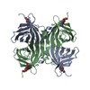
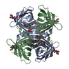
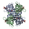
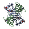
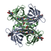
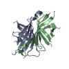
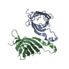
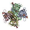
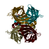
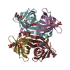
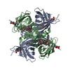
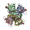
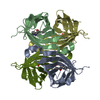
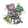
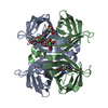
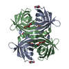
SHEET DETERMINATION METHOD: DSSP THE SHEETS PRESENTED AS "AA BA" IN EACH CHAIN ON SHEET RECORDS ... SHEET DETERMINATION METHOD: DSSP THE SHEETS PRESENTED AS "AA BA" IN EACH CHAIN ON SHEET RECORDS BELOW IS ACTUALLY AN 9-STRANDED BARREL THIS IS REPRESENTED BY A 10-STRANDED SHEET IN WHICH THE FIRST AND LAST STRANDS ARE IDENTICAL.

-
Structure visualization

Structure viewerMolecule:
MolmilJmol/JSmol

Downloads & links

-
Assembly

Deposited unit
A: AVIDIN
B: AVIDIN
hetero molecules


Theoretical massNumber of molelcules
Total (without water)29,2758
Polymers28,7222
Non-polymers5536
Water2,684149
1
A: AVIDIN
B: AVIDIN
hetero molecules

A: AVIDIN
B: AVIDIN
hetero molecules


Theoretical massNumber of molelcules
Total (without water)58,55016
Polymers57,4454
Non-polymers1,10512
Water724
TypeNameSymmetry operationNumber
identity operation1_555x,y,z1
crystal symmetry operation2_555-x,-y,z1
Buried area12110 Å2
ΔGint-58.1 kcal/mol
Surface area23550 Å2
MethodPQS
Unit cell
Length a, b, c (Å)72.949, 78.772, 43.010
Angle α, β, γ (deg.)90.00, 90.00, 90.00
Int Tables number18
Space group name H-MP21212
Noncrystallographic symmetry (NCS)NCS oper: (Code: given
Matrix: (-0.2391, 0.971, 0.0016), (0.971, 0.2391, -0.0037), (-0.004, 0.0007, -1)
Vector: 0.0169, 0.0719, 14.8821)

-
Components

#1: Protein AVIDIN


Mass: 14361.149 Da / Num. of mol.: 2
Source method: isolated from a genetically manipulated source
Source: (gene. exp.) GALLUS GALLUS (chicken) / Production host: ESCHERICHIA COLI (E. coli) / References: UniProt: P02701
#2: Chemical
ChemComp-GOL / GLYCEROL / GLYCERIN / PROPANE-1,2,3-TRIOL


Mass: 92.094 Da / Num. of mol.: 6 / Source method: obtained synthetically / Formula: C3H8O3
#3: Water ChemComp-HOH / water


Mass: 18.015 Da / Num. of mol.: 149 / Source method: isolated from a natural source / Formula: H2O
Compound detailsFUNCTION: THE BIOLOGICAL FUNCTION OF AVIDIN IS NOT KNOWN. FORMS A STRONG NON-COVALENT SPECIFIC ...FUNCTION: THE BIOLOGICAL FUNCTION OF AVIDIN IS NOT KNOWN. FORMS A STRONG NON-COVALENT SPECIFIC COMPLEX WITH BIOTIN (ONE MOLECULE OF BIOTIN PER SUBUNIT OF AVIDIN).

-
Experimental details

-
Experiment

ExperimentMethod: X-RAY DIFFRACTION / Number of used crystals: 1

-
Sample preparation

CrystalDensity Matthews: 2.15 Å3/Da / Density % sol: 42.82 %
Crystal growTemperature: 295 K / Method: vapor diffusion, hanging drop / pH: 6.6
Details: Equal volumes (1 ul) of protein (0.5 mg/ml) in 50 mM Na acetate (pH 4) + 20 mM NaCl and well solution of 0.1 M MES (pH 6.6) + 24% PEG 8000 + 0.2 M Mg acetate

-
Data collection

DiffractionMean temperature: 100 K
Diffraction sourceSource: SYNCHROTRON / Site: EMBL/DESY, HAMBURG / Beamline: X13 / Wavelength: 0.804
DetectorType: MARRESEARCH / Detector: CCD
RadiationProtocol: SINGLE WAVELENGTH / Monochromatic (M) / Laue (L): M / Scattering type: x-ray
Radiation wavelengthWavelength: 0.804 Å / Relative weight: 1
ReflectionResolution: 1.48→20 Å / Num. obs: 41811 / % possible obs: 99 % / Redundancy: 4 % / Rmerge(I) obs: 0.073 / Net I/σ(I): 10
Reflection shellResolution: 1.48→1.58 Å / Redundancy: 4 % / Rmerge(I) obs: 0.47 / Mean I/σ(I) obs: 3 / % possible all: 99

-
Processing

Software
NameVersionClassification
REFMAC5.1.24refinement
XDSdata reduction
XDSdata scaling
AMoREphasing
RefinementMethod to determine structure: MOLECULAR REPLACEMENT
Starting model: PDB ENTRY 1AVD
Resolution: 1.48→19.1 Å / Cor.coef. Fo:Fc: 0.964 / Cor.coef. Fo:Fc free: 0.955 / SU B: 1.213 / SU ML: 0.045 / Cross valid method: THROUGHOUT / ESU R: 0.067 / ESU R Free: 0.068 / Stereochemistry target values: MAXIMUM LIKELIHOOD
Details: HYDROGENS HAVE BEEN ADDED IN THE RIDING POSITIONS. THE PEPTIDE BONDS BETWEEN RESIDUES A41 AND A42 AND BETWEEN B41 AND B42 HAVE BEEN DETERMINED TO BE CIS- BONDS. THESE RESIDUES ARE NOT ...Details: HYDROGENS HAVE BEEN ADDED IN THE RIDING POSITIONS. THE PEPTIDE BONDS BETWEEN RESIDUES A41 AND A42 AND BETWEEN B41 AND B42 HAVE BEEN DETERMINED TO BE CIS- BONDS. THESE RESIDUES ARE NOT EXPECTED TO ADOPT THE CIS- CONFORMATION. THIS IS DUE TO THE POOR ELECTRON DENSITY IN THESE REGIONS OF THE ELECTRON DENSITY MAP. RESIDUES 34-46 OF BOTH CHAINS FORM LOOPS WITH TWO ALTERNATE CONFORMATIONS. BOTH ALTERNATE CONFORMATIONS CAN BE DETERMINED FOR RESIDUES 34-36 AND 42-46, BUT DUE TO POOR ELECTRON DENSITY RESIDUES 37-41 CAN BE ASSIGNED ONLY A SINGLE CONFORMATION.
RfactorNum. reflection% reflectionSelection details
Rfree0.19 2091 5 %RANDOM
Rwork0.165 ---
obs0.167 39720 100 %-
Solvent computationIon probe radii: 0.8 Å / Shrinkage radii: 0.8 Å / VDW probe radii: 1.4 Å / Solvent model: BABINET MODEL WITH MASK
Displacement parametersBiso mean: 7.58 Å2
Baniso -1Baniso -2Baniso -3
1-1.13 Å20 Å20 Å2
2---0.35 Å20 Å2
3----0.77 Å2
Refinement stepCycle: LAST / Resolution: 1.48→19.1 Å
ProteinNucleic acidLigandSolventTotal
Num. atoms1905 0 36 149 2090
Refine LS restraints
Refine-IDTypeDev idealDev ideal targetNumber
X-RAY DIFFRACTIONr_bond_refined_d0.0130.0212134
X-RAY DIFFRACTIONr_bond_other_d0.0030.021914
X-RAY DIFFRACTIONr_angle_refined_deg1.4831.9242887
X-RAY DIFFRACTIONr_angle_other_deg0.84934477
X-RAY DIFFRACTIONr_dihedral_angle_1_deg6.9725257
X-RAY DIFFRACTIONr_dihedral_angle_2_deg
X-RAY DIFFRACTIONr_dihedral_angle_3_deg
X-RAY DIFFRACTIONr_dihedral_angle_4_deg
X-RAY DIFFRACTIONr_chiral_restr0.0970.2343
X-RAY DIFFRACTIONr_gen_planes_refined0.0060.022290
X-RAY DIFFRACTIONr_gen_planes_other0.0020.02427
X-RAY DIFFRACTIONr_nbd_refined0.1920.2373
X-RAY DIFFRACTIONr_nbd_other0.2570.22260
X-RAY DIFFRACTIONr_nbtor_refined
X-RAY DIFFRACTIONr_nbtor_other0.0880.21330
X-RAY DIFFRACTIONr_xyhbond_nbd_refined0.1490.296
X-RAY DIFFRACTIONr_xyhbond_nbd_other
X-RAY DIFFRACTIONr_metal_ion_refined
X-RAY DIFFRACTIONr_metal_ion_other
X-RAY DIFFRACTIONr_symmetry_vdw_refined0.2470.249
X-RAY DIFFRACTIONr_symmetry_vdw_other0.3030.2197
X-RAY DIFFRACTIONr_symmetry_hbond_refined0.1410.222
X-RAY DIFFRACTIONr_symmetry_hbond_other
X-RAY DIFFRACTIONr_symmetry_metal_ion_refined
X-RAY DIFFRACTIONr_symmetry_metal_ion_other
X-RAY DIFFRACTIONr_mcbond_it0.71.51285
X-RAY DIFFRACTIONr_mcbond_other
X-RAY DIFFRACTIONr_mcangle_it1.22622114
X-RAY DIFFRACTIONr_mcangle_other
X-RAY DIFFRACTIONr_scbond_it2.0133849
X-RAY DIFFRACTIONr_scbond_other
X-RAY DIFFRACTIONr_scangle_it3.2464.5773
X-RAY DIFFRACTIONr_scangle_other
X-RAY DIFFRACTIONr_long_range_B_refined
X-RAY DIFFRACTIONr_long_range_B_other
X-RAY DIFFRACTIONr_rigid_bond_restr
X-RAY DIFFRACTIONr_sphericity_free
X-RAY DIFFRACTIONr_sphericity_bonded
LS refinement shellResolution: 1.48→1.52 Å / Total num. of bins used: 20 /
RfactorNum. reflection
Rfree0.286 152
Rwork0.254 2878
Refinement TLS params.

Method: refined / Refine-ID: X-RAY DIFFRACTION

IDL112)L122)L132)L222)L232)L332)S11 (Å °)S12 (Å °)S13 (Å °)S21 (Å °)S22 (Å °)S23 (Å °)S31 (Å °)S32 (Å °)S33 (Å °)T112)T122)T132)T222)T232)T332)Origin x (Å)Origin y (Å)Origin z (Å)
12.7193-1.07471.85857.32884.24087.92310.12450.2604-0.158-0.6367-0.06830.0551-0.0939-0.1316-0.05620.1457-0.0046-0.00040.18110.04020.1539-3.45416.029-8.855
22.7157-0.6074-2.0201-2.3362-0.82519.12220.0724-0.2556-0.1887-0.04430.08220.3024-0.0972-0.2961-0.15460.314-0.049-0.01160.27340.01370.282516.6310.30823.692
32.824-0.7272-0.65852.4720.16042.3247-0.0537-0.13290.17320.24740.0376-0.085-0.16890.04350.01610.1190.0169-0.0170.0919-0.00070.1117-7.89921.0988.321
42.41310.02820.29733.2377-0.78741.902-0.01310.11850.0345-0.0093-0.0988-0.269-0.08770.26240.11190.0323-0.0155-0.02180.17820.02590.146522.608-2.1565.725
51.1466-0.11270.30491.2939-0.04953.283-0.0388-0.02260.10760.1080.0334-0.0002-0.3145-0.04910.00540.03730.01770.0010.04580.00350.067-8.76513.988.801
662.416548.467.495978.852-38.012245.6776-0.1495-0.2498-1.4287-0.0179-0.3422-2.36010.64041.61840.49170.3773-0.0462-0.00020.3438-0.02240.34086.74822.81118.486
72.18870.98890.61024.30451.11121.8954-0.01520.11550.041-0.04750.0201-0.0694-0.11140.059-0.00490.00270.00930.00860.06220.01410.0351-2.8286.7082.494
81.2312-0.1023-0.33271.3649-0.32091.8257-0.0169-0.01820.06230.09940.011-0.0798-0.07260.14130.00590.0184-0.0042-0.01880.0616-0.00320.061612.239-2.0948.076
Refinement TLS group
IDRefine-IDRefine TLS-IDAuth asym-IDAuth seq-ID
1X-RAY DIFFRACTION1A34 - 45
2X-RAY DIFFRACTION2B34 - 45
3X-RAY DIFFRACTION3A3 - 33
4X-RAY DIFFRACTION4B2 - 33
5X-RAY DIFFRACTION5A46 - 84
6X-RAY DIFFRACTION6A85 - 90
7X-RAY DIFFRACTION7A91 - 123
8X-RAY DIFFRACTION8B46 - 123

+
About Yorodumi

-
News

-
Feb 9, 2022. New format data for meta-information of EMDB entries

New format data for meta-information of EMDB entries

  • Version 3 of the EMDB header file is now the official format.
  • The previous official version 1.9 will be removed from the archive.

Related info.:EMDB header

External links:wwPDB to switch to version 3 of the EMDB data model

-
Aug 12, 2020. Covid-19 info

Covid-19 info

URL: https://pdbjlvh1.pdbj.org/emnavi/covid19.php

New page: Covid-19 featured information page in EM Navigator.

Related info.:Covid-19 info / Mar 5, 2020. Novel coronavirus structure data

+
Mar 5, 2020. Novel coronavirus structure data

Novel coronavirus structure data

Related info.:Yorodumi Speices / Aug 12, 2020. Covid-19 info

External links:COVID-19 featured content - PDBj / Molecule of the Month (242):Coronavirus Proteases

+
Jan 31, 2019. EMDB accession codes are about to change! (news from PDBe EMDB page)

EMDB accession codes are about to change! (news from PDBe EMDB page)

  • The allocation of 4 digits for EMDB accession codes will soon come to an end. Whilst these codes will remain in use, new EMDB accession codes will include an additional digit and will expand incrementally as the available range of codes is exhausted. The current 4-digit format prefixed with “EMD-” (i.e. EMD-XXXX) will advance to a 5-digit format (i.e. EMD-XXXXX), and so on. It is currently estimated that the 4-digit codes will be depleted around Spring 2019, at which point the 5-digit format will come into force.
  • The EM Navigator/Yorodumi systems omit the EMD- prefix.

Related info.:Q: What is EMD? / ID/Accession-code notation in Yorodumi/EM Navigator

External links:EMDB Accession Codes are Changing Soon! / Contact to PDBj

+
Jul 12, 2017. Major update of PDB

Major update of PDB

  • wwPDB released updated PDB data conforming to the new PDBx/mmCIF dictionary.
  • This is a major update changing the version number from 4 to 5, and with Remediation, in which all the entries are updated.
  • In this update, many items about electron microscopy experimental information are reorganized (e.g. em_software).
  • Now, EM Navigator and Yorodumi are based on the updated data.

External links:wwPDB Remediation / Enriched Model Files Conforming to OneDep Data Standards Now Available in the PDB FTP Archive

-
Yorodumi

Thousand views of thousand structures

  • Yorodumi is a browser for structure data from EMDB, PDB, SASBDB, etc.
  • This page is also the successor to EM Navigator detail page, and also detail information page/front-end page for Omokage search.
  • The word "yorodu" (or yorozu) is an old Japanese word meaning "ten thousand". "mi" (miru) is to see.

Related info.:EMDB / PDB / SASBDB / Comparison of 3 databanks / Yorodumi Search / Aug 31, 2016. New EM Navigator & Yorodumi / Yorodumi Papers / Jmol/JSmol / Function and homology information / Changes in new EM Navigator and Yorodumi

Read more