 Movie
Movie Controller


+ Open data

- Basic information
| Entry | Database: PDB / ID: 1rav | ||||||
|---|---|---|---|---|---|---|---|
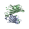
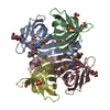
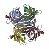
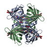
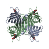
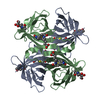
| Title | RECOMBINANT AVIDIN | ||||||
 Components Components | AVIDIN | ||||||
 Keywords Keywords | GLYCOPROTEIN / AVIDIN / BIOTIN BINDING PROTEIN / CALYCINS / UP-AND-DOWN BETA BARREL | ||||||
| Function / homology |  Function and homology information Function and homology information | ||||||
| Biological species |  | ||||||
| Method |  X-RAY DIFFRACTION / MOLECULAR REPLACEMENT / Resolution: 2.2 Å X-RAY DIFFRACTION / MOLECULAR REPLACEMENT / Resolution: 2.2 Å | ||||||
 Authors Authors | Rosano, C. / Arosio, P. / Bolognesi, M. | ||||||
 Citation Citation | Journal: Eur.J.Biochem. / Year: 1998 Title: Biochemical characterization and crystal structure of a recombinant hen avidin and its acidic mutant expressed in Escherichia coli. Authors: Nardone, E. / Rosano, C. / Santambrogio, P. / Curnis, F. / Corti, A. / Magni, F. / Siccardi, A.G. / Paganelli, G. / Losso, R. / Apreda, B. / Bolognesi, M. / Sidoli, A. / Arosio, P. #1: Journal: J.Mol.Biol. / Year: 1994Title: Crystal Structure of Apo-Avidin from Hen Egg-White Authors: Pugliese, L. / Malcovati, M. / Coda, A. / Bolognesi, M. #2: Journal: Proc.Natl.Acad.Sci.USA / Year: 1993Title: Three-Dimensional Structures of Avidin and the Avidin-Biotin Complex Authors: Livnah, O. / Bayer, E.A. / Wilchek, M. / Sussman, J.L. #3: Journal: J.Mol.Biol. / Year: 1993Title: Three-Dimensional Structure of the Tetragonal Crystal Form of Egg-White Avidin in its Functional Complex with Biotin at 2.7 A Resolution Authors: Pugliese, L. / Coda, A. / Malcovati, M. / Bolognesi, M. | ||||||
| History |
|
- Structure visualization
| Structure viewer | Molecule: MolmilJmol/JSmol |
|---|
- Downloads & links
-Download
| PDBx/mmCIF format | 1rav.cif.gz | 64.4 KB | Display | PDBx/mmCIF format |
|---|---|---|---|---|
| PDB format | pdb1rav.ent.gz | 46.7 KB | Display | PDB format |
| PDBx/mmJSON format | 1rav.json.gz | Tree view | PDBx/mmJSON format | |
| Others |  Other downloads Other downloads |
-Validation report
| Arichive directory | https://data.pdbj.org/pub/pdb/validation_reports/ra/1ravftp://data.pdbj.org/pub/pdb/validation_reports/ra/1rav | HTTPS FTP |
|---|
-Related structure data
| Related structure data | 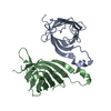 2camC  1aveS S: Starting model for refinement C: citing same article ( |
|---|---|
| Similar structure data |


-Links
 PDBj
PDBj- Assembly
| Deposited unit | 
| ||||||||
|---|---|---|---|---|---|---|---|---|---|
| 1 | 
| ||||||||
| Unit cell |
|
-Components
| #1: Protein | Mass: 14291.058 Da / Num. of mol.: 2 Source method: isolated from a genetically manipulated source Source: (gene. exp.)  #2: Water | ChemComp-HOH / |  Mass: 18.015 Da / Num. of mol.: 80 / Source method: isolated from a natural source / Formula: H2O Mass: 18.015 Da / Num. of mol.: 80 / Source method: isolated from a natural source / Formula: H2OHas protein modification | Y | |
|---|
-Experimental details
-Experiment
| Experiment | Method: X-RAY DIFFRACTION / Number of used crystals: 1 |
|---|
- Sample preparation
| Crystal | Density Matthews: 2.42 Å3/Da / Density % sol: 58.7 % | ||||||||||||||||||||||||||||||||||||
|---|---|---|---|---|---|---|---|---|---|---|---|---|---|---|---|---|---|---|---|---|---|---|---|---|---|---|---|---|---|---|---|---|---|---|---|---|---|
| Crystal grow | Method: vapor diffusion / pH: 7.2 Details: RECOMBINANT AVIDIN WAS CRYSTALLIZED FROM 9% W/V PEG 8000. PH 7.2 0.05 M TRIS BUFFER AT 22 C BY VAPOUR DIFFUSION TECHNIQUES., vapor diffusion | ||||||||||||||||||||||||||||||||||||
| Crystal | *PLUS | ||||||||||||||||||||||||||||||||||||
| Crystal grow | *PLUS Temperature: 22 ℃ / pH: 7.2 / Method: vapor diffusion | ||||||||||||||||||||||||||||||||||||
| Components of the solutions | *PLUS
|
-Data collection
| Diffraction | Mean temperature: 293 K |
|---|---|
| Diffraction source | Source: ROTATING ANODE / Type: RIGAKU RUH2R / Wavelength: 1.5418 |
| Detector | Type: RIGAKU RAXIS IIC / Detector: IMAGE PLATE / Date: Oct 1, 1997 |
| Radiation | Monochromator: GRAPHITE(002) / Monochromatic (M) / Laue (L): M / Scattering type: x-ray |
| Radiation wavelength | Wavelength: 1.5418 Å / Relative weight: 1 |
| Reflection | Resolution: 2.2→20.5 Å / Num. obs: 13827 / % possible obs: 95 % / Observed criterion σ(I): 0 / Redundancy: 3.6 % / Rmerge(I) obs: 0.054 / Net I/σ(I): 15 |
| Reflection shell | Resolution: 2.2→2.7 Å / % possible all: 93.1 |
| Reflection | *PLUS Num. measured all: 48932 |
- Processing
| Software |
| ||||||||||||||||||||||||||||||||||||||||||||||||||
|---|---|---|---|---|---|---|---|---|---|---|---|---|---|---|---|---|---|---|---|---|---|---|---|---|---|---|---|---|---|---|---|---|---|---|---|---|---|---|---|---|---|---|---|---|---|---|---|---|---|---|---|
| Refinement | Method to determine structure: MOLECULAR REPLACEMENT Starting model: PDB ENTRY 1AVE Resolution: 2.2→20.2 Å / Isotropic thermal model: TNT BCORREL / σ(F): 0 / Stereochemistry target values: TNT PROTGEO
| ||||||||||||||||||||||||||||||||||||||||||||||||||
| Solvent computation | Solvent model: TNT / Bsol: 150 Å2 / ksol: 0.77 e/Å3 | ||||||||||||||||||||||||||||||||||||||||||||||||||
| Refinement step | Cycle: LAST / Resolution: 2.2→20.2 Å
| ||||||||||||||||||||||||||||||||||||||||||||||||||
| Refine LS restraints |
| ||||||||||||||||||||||||||||||||||||||||||||||||||
| Software | *PLUS Name: TNT / Version: 5E / Classification: refinement | ||||||||||||||||||||||||||||||||||||||||||||||||||
| Refinement | *PLUS Rfactor all: 0.17 | ||||||||||||||||||||||||||||||||||||||||||||||||||
| Solvent computation | *PLUS | ||||||||||||||||||||||||||||||||||||||||||||||||||
| Displacement parameters | *PLUS | ||||||||||||||||||||||||||||||||||||||||||||||||||
| Refine LS restraints | *PLUS
|
