 Movie
Movie Controller


+ Open data

- Basic information
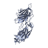
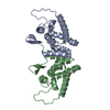
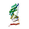
| Entry | Database: PDB / ID: 1vpp | ||||||
|---|---|---|---|---|---|---|---|
| Title | COMPLEX BETWEEN VEGF AND A RECEPTOR BLOCKING PEPTIDE | ||||||
 Components Components |
| ||||||
 Keywords Keywords | GROWTH FACTOR/GROWTH FACTOR INHIBITOR / CYSTINE KNOT / ANGIOGENESIS / VASCULOGENESIS / RECEPTOR BLOCKING PEPTIDE / GROWTH FACTOR-GROWTH FACTOR INHIBITOR COMPLEX | ||||||
| Function / homology |  Function and homology information Function and homology informationSignaling by VEGF / basophil chemotaxis / positive regulation of endothelial cell chemotaxis by VEGF-activated vascular endothelial growth factor receptor signaling pathway / VEGF-A complex / cellular stress response to acid chemical / positive regulation of lymphangiogenesis / VEGF ligand-receptor interactions / vascular endothelial growth factor receptor 1 binding / negative regulation of adherens junction organization / primitive erythrocyte differentiation ...Signaling by VEGF / basophil chemotaxis / positive regulation of endothelial cell chemotaxis by VEGF-activated vascular endothelial growth factor receptor signaling pathway / VEGF-A complex / cellular stress response to acid chemical / positive regulation of lymphangiogenesis / VEGF ligand-receptor interactions / vascular endothelial growth factor receptor 1 binding / negative regulation of adherens junction organization / primitive erythrocyte differentiation / positive regulation of mast cell chemotaxis / negative regulation of establishment of endothelial barrier / vascular endothelial growth factor receptor binding / post-embryonic camera-type eye development / lymph vessel morphogenesis / negative regulation of blood-brain barrier permeability / positive regulation of cell proliferation by VEGF-activated platelet derived growth factor receptor signaling pathway / : / VEGF-activated neuropilin signaling pathway / bone trabecula formation / coronary vein morphogenesis / cardiac vascular smooth muscle cell development / lymphangiogenesis / motor neuron migration / VEGF binds to VEGFR leading to receptor dimerization / vascular endothelial growth factor receptor-2 signaling pathway / eye photoreceptor cell development / lung vasculature development / regulation of hematopoietic progenitor cell differentiation / positive regulation of axon extension involved in axon guidance / endothelial cell chemotaxis / positive regulation of protein localization to early endosome / positive regulation of protein autophosphorylation / positive regulation of trophoblast cell migration / vascular wound healing / positive regulation of epithelial tube formation / camera-type eye morphogenesis / neuropilin binding / positive regulation of blood vessel endothelial cell proliferation involved in sprouting angiogenesis / positive regulation of branching involved in ureteric bud morphogenesis / induction of positive chemotaxis / transmembrane receptor protein tyrosine kinase activator activity / coronary artery morphogenesis / negative regulation of cell-cell adhesion mediated by cadherin / tube formation / vascular endothelial growth factor receptor 2 binding / dopaminergic neuron differentiation / positive regulation of vascular permeability / commissural neuron axon guidance / positive regulation of vascular endothelial growth factor signaling pathway / cell migration involved in sprouting angiogenesis / positive regulation of peptidyl-tyrosine phosphorylation / positive regulation of blood vessel branching / surfactant homeostasis / platelet-derived growth factor receptor binding / epithelial cell maturation / sprouting angiogenesis / extracellular matrix binding / endothelial cell proliferation / positive regulation of positive chemotaxis / Regulation of gene expression by Hypoxia-inducible Factor / positive regulation of leukocyte migration / artery morphogenesis / retinal ganglion cell axon guidance / positive regulation of endothelial cell chemotaxis / cardiac muscle cell development / positive regulation of cell migration involved in sprouting angiogenesis / positive regulation of DNA biosynthetic process / negative regulation of epithelial to mesenchymal transition / vascular endothelial growth factor signaling pathway / branching involved in blood vessel morphogenesis / positive regulation of neuroblast proliferation / positive chemotaxis / negative regulation of fat cell differentiation / positive regulation of sprouting angiogenesis / chemoattractant activity / mesoderm development / outflow tract morphogenesis / fibronectin binding / positive regulation of cell division / macrophage differentiation / neuroblast proliferation / mammary gland alveolus development / cellular response to vascular endothelial growth factor stimulus / positive regulation of receptor internalization / positive regulation of focal adhesion assembly / monocyte differentiation / positive regulation of blood vessel endothelial cell migration / vascular endothelial growth factor receptor signaling pathway / positive regulation of osteoblast differentiation / vasculogenesis / ovarian follicle development / heart morphogenesis / lactation / TFAP2 (AP-2) family regulates transcription of growth factors and their receptors / cell maturation / positive regulation of endothelial cell proliferation / epithelial cell differentiation / lung development / positive regulation of endothelial cell migration Similarity search - Function | ||||||
| Biological species |  Homo sapiens (human) Homo sapiens (human) | ||||||
| Method |  X-RAY DIFFRACTION / SYNCHROTRON / MOLECULAR REPLACEMENT / Resolution: 1.9 Å X-RAY DIFFRACTION / SYNCHROTRON / MOLECULAR REPLACEMENT / Resolution: 1.9 Å | ||||||
 Authors Authors | Wiesmann, C. / Christinger, H.W. / Cochran, A.G. / Cunningham, B.C. / Fairbrother, W.J. / Keenan, C.J. / Meng, G. / de Vos, A.M. | ||||||
 Citation Citation | Journal: Biochemistry / Year: 1998 Title: Crystal structure of the complex between VEGF and a receptor-blocking peptide. Authors: Wiesmann, C. / Christinger, H.W. / Cochran, A.G. / Cunningham, B.C. / Fairbrother, W.J. / Keenan, C.J. / Meng, G. / de Vos, A.M. | ||||||
| History |
|
- Structure visualization
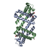
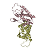
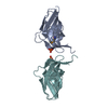
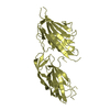
| Structure viewer | Molecule: MolmilJmol/JSmol |
|---|
- Downloads & links
-Download
| PDBx/mmCIF format | 1vpp.cif.gz | 66.8 KB | Display | PDBx/mmCIF format |
|---|---|---|---|---|
| PDB format | pdb1vpp.ent.gz | 48.7 KB | Display | PDB format |
| PDBx/mmJSON format | 1vpp.json.gz | Tree view | PDBx/mmJSON format | |
| Others |  Other downloads Other downloads |
-Validation report
| Arichive directory | https://data.pdbj.org/pub/pdb/validation_reports/vp/1vppftp://data.pdbj.org/pub/pdb/validation_reports/vp/1vpp | HTTPS FTP |
|---|
-Related structure data
| Related structure data | 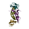 1fltS S: Starting model for refinement |
|---|---|
| Similar structure data |


-Links
 PDBj
PDBj


- Assembly
| Deposited unit | 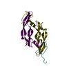
| ||||||||
|---|---|---|---|---|---|---|---|---|---|
| 1 |
| ||||||||
| Unit cell |
| ||||||||
| Noncrystallographic symmetry (NCS) | NCS oper: (Code: given Matrix: (0.48459, 0.84119, -0.23993), Vector: |
-Components
| #1: Protein | Mass: 11948.680 Da / Num. of mol.: 2 / Fragment: RECEPTOR BINDING DOMAIN Source method: isolated from a genetically manipulated source Source: (gene. exp.) Homo sapiens (human) / Production host:  #2: Protein/peptide | Mass: 2258.491 Da / Num. of mol.: 2 / Fragment: RECEPTOR BLOCKING PEPTIDE / Source method: obtained synthetically / Details: THIS SEQUENCE IS CHEMICALLY SYNTHESIZED #3: Water | ChemComp-HOH / |  Mass: 18.015 Da / Num. of mol.: 383 / Source method: isolated from a natural source / Formula: H2O Mass: 18.015 Da / Num. of mol.: 383 / Source method: isolated from a natural source / Formula: H2OHas protein modification | Y | |
|---|
-Experimental details
-Experiment
| Experiment | Method: X-RAY DIFFRACTION / Number of used crystals: 1 |
|---|
- Sample preparation
| Crystal | Density Matthews: 2.5 Å3/Da / Density % sol: 50.84 % | ||||||||||||||||||||||||||||||
|---|---|---|---|---|---|---|---|---|---|---|---|---|---|---|---|---|---|---|---|---|---|---|---|---|---|---|---|---|---|---|---|
| Crystal grow | pH: 7.5 / Details: pH 7.5 | ||||||||||||||||||||||||||||||
| Crystal grow | *PLUS Method: vapor diffusion, hanging drop / pH: 5 | ||||||||||||||||||||||||||||||
| Components of the solutions | *PLUS
|
-Data collection
| Diffraction | Mean temperature: 100 K |
|---|---|
| Diffraction source | Source: SYNCHROTRON / Site: SSRL  / Beamline: BL7-1 / Wavelength: 1.08 / Beamline: BL7-1 / Wavelength: 1.08 |
| Detector | Type: MARRESEARCH / Detector: IMAGE PLATE / Date: May 15, 1997 |
| Radiation | Protocol: SINGLE WAVELENGTH / Monochromatic (M) / Laue (L): M / Scattering type: x-ray |
| Radiation wavelength | Wavelength: 1.08 Å / Relative weight: 1 |
| Reflection | Resolution: 1.9→99 Å / Num. obs: 23253 / % possible obs: 99.6 % / Redundancy: 5.1 % / Rsym value: 0.06 / Net I/σ(I): 14.4 |
| Reflection | *PLUS Rmerge(I) obs: 0.045 |
- Processing
| Software |
| ||||||||||||||||||||||||||||||||||||||||||||||||||||||||||||||||||||||||||||||||||||
|---|---|---|---|---|---|---|---|---|---|---|---|---|---|---|---|---|---|---|---|---|---|---|---|---|---|---|---|---|---|---|---|---|---|---|---|---|---|---|---|---|---|---|---|---|---|---|---|---|---|---|---|---|---|---|---|---|---|---|---|---|---|---|---|---|---|---|---|---|---|---|---|---|---|---|---|---|---|---|---|---|---|---|---|---|---|
| Refinement | Method to determine structure: MOLECULAR REPLACEMENT Starting model: 1FLT Resolution: 1.9→20 Å / Cross valid method: THROUGHOUT / σ(F): 0 / ESU R: 0.15 / ESU R Free: 0.17
| ||||||||||||||||||||||||||||||||||||||||||||||||||||||||||||||||||||||||||||||||||||
| Displacement parameters | Biso mean: 27.1 Å2 /
| ||||||||||||||||||||||||||||||||||||||||||||||||||||||||||||||||||||||||||||||||||||
| Refinement step | Cycle: LAST / Resolution: 1.9→20 Å
| ||||||||||||||||||||||||||||||||||||||||||||||||||||||||||||||||||||||||||||||||||||
| Refine LS restraints |
| ||||||||||||||||||||||||||||||||||||||||||||||||||||||||||||||||||||||||||||||||||||
| Software | *PLUS Name: REFMAC / Classification: refinement | ||||||||||||||||||||||||||||||||||||||||||||||||||||||||||||||||||||||||||||||||||||
| Refinement | *PLUS Highest resolution: 1.9 Å / Num. reflection obs: 21270 / σ(F): 0 / % reflection Rfree: 6.3 % / Rfactor obs: 0.19 / Rfactor Rfree: 0.27 / Rfactor Rwork: 0.19 | ||||||||||||||||||||||||||||||||||||||||||||||||||||||||||||||||||||||||||||||||||||
| Solvent computation | *PLUS | ||||||||||||||||||||||||||||||||||||||||||||||||||||||||||||||||||||||||||||||||||||
| Displacement parameters | *PLUS Biso mean: 27.1 Å2 | ||||||||||||||||||||||||||||||||||||||||||||||||||||||||||||||||||||||||||||||||||||
| Refine LS restraints | *PLUS
|
