 Movie
Movie Controller


+ Open data

- Basic information
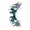
| Entry | Database: PDB / ID: 1flt | ||||||
|---|---|---|---|---|---|---|---|
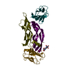
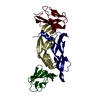
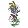
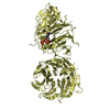
| Title | VEGF IN COMPLEX WITH DOMAIN 2 OF THE FLT-1 RECEPTOR | ||||||
 Components Components |
| ||||||
 Keywords Keywords | COMPLEX (GROWTH FACTOR/TRANSFERASE) / COMPLEX (GROWTH FACTOR-TRANSFERASE) / FLT-1 RECEPTOR / CYSTINE KNOT / GLYCOPROTEIN / IMMUNOGLOBULIN-LIKE DOMAIN TRANSFERASE / COMPLEX (GROWTH FACTOR-TRANSFERASE) complex | ||||||
| Function / homology |  Function and homology information Function and homology informationvascular endothelial growth factor receptor-1 signaling pathway / placental growth factor receptor activity / Signaling by VEGF / basophil chemotaxis / positive regulation of endothelial cell chemotaxis by VEGF-activated vascular endothelial growth factor receptor signaling pathway / VEGF-A complex / cellular stress response to acid chemical / positive regulation of lymphangiogenesis / VEGF ligand-receptor interactions / vascular endothelial growth factor receptor 1 binding ...vascular endothelial growth factor receptor-1 signaling pathway / placental growth factor receptor activity / Signaling by VEGF / basophil chemotaxis / positive regulation of endothelial cell chemotaxis by VEGF-activated vascular endothelial growth factor receptor signaling pathway / VEGF-A complex / cellular stress response to acid chemical / positive regulation of lymphangiogenesis / VEGF ligand-receptor interactions / vascular endothelial growth factor receptor 1 binding / negative regulation of adherens junction organization / primitive erythrocyte differentiation / positive regulation of mast cell chemotaxis / negative regulation of establishment of endothelial barrier / vascular endothelial growth factor receptor binding / post-embryonic camera-type eye development / lymph vessel morphogenesis / negative regulation of blood-brain barrier permeability / positive regulation of cell proliferation by VEGF-activated platelet derived growth factor receptor signaling pathway / : / VEGF-activated neuropilin signaling pathway / bone trabecula formation / hyaloid vascular plexus regression / coronary vein morphogenesis / cardiac vascular smooth muscle cell development / lymphangiogenesis / Neuropilin interactions with VEGF and VEGFR / motor neuron migration / VEGF binds to VEGFR leading to receptor dimerization / vascular endothelial growth factor receptor-2 signaling pathway / eye photoreceptor cell development / lung vasculature development / regulation of hematopoietic progenitor cell differentiation / vascular endothelial growth factor receptor activity / positive regulation of axon extension involved in axon guidance / endothelial cell chemotaxis / positive regulation of protein localization to early endosome / positive regulation of protein autophosphorylation / positive regulation of trophoblast cell migration / vascular wound healing / embryonic morphogenesis / positive regulation of epithelial tube formation / camera-type eye morphogenesis / neuropilin binding / positive regulation of blood vessel endothelial cell proliferation involved in sprouting angiogenesis / positive regulation of branching involved in ureteric bud morphogenesis / induction of positive chemotaxis / transmembrane receptor protein tyrosine kinase activator activity / coronary artery morphogenesis / negative regulation of cell-cell adhesion mediated by cadherin / tube formation / vascular endothelial growth factor receptor 2 binding / dopaminergic neuron differentiation / positive regulation of vascular permeability / commissural neuron axon guidance / positive regulation of vascular endothelial growth factor signaling pathway / cell migration involved in sprouting angiogenesis / positive regulation of peptidyl-tyrosine phosphorylation / positive regulation of blood vessel branching / surfactant homeostasis / platelet-derived growth factor receptor binding / epithelial cell maturation / sprouting angiogenesis / extracellular matrix binding / endothelial cell proliferation / blood vessel morphogenesis / positive regulation of positive chemotaxis / Regulation of gene expression by Hypoxia-inducible Factor / negative regulation of vascular endothelial cell proliferation / positive regulation of leukocyte migration / artery morphogenesis / retinal ganglion cell axon guidance / positive regulation of endothelial cell chemotaxis / cardiac muscle cell development / positive regulation of cell migration involved in sprouting angiogenesis / positive regulation of DNA biosynthetic process / negative regulation of epithelial to mesenchymal transition / vascular endothelial growth factor signaling pathway / branching involved in blood vessel morphogenesis / positive regulation of neuroblast proliferation / growth factor binding / positive chemotaxis / negative regulation of fat cell differentiation / positive regulation of sprouting angiogenesis / chemoattractant activity / positive regulation of MAP kinase activity / mesoderm development / outflow tract morphogenesis / fibronectin binding / positive regulation of cell division / macrophage differentiation / neuroblast proliferation / mammary gland alveolus development / cellular response to vascular endothelial growth factor stimulus / positive regulation of receptor internalization / monocyte chemotaxis / positive regulation of focal adhesion assembly / monocyte differentiation / positive regulation of blood vessel endothelial cell migration / vascular endothelial growth factor receptor signaling pathway Similarity search - Function | ||||||
| Biological species |  Homo sapiens (human) Homo sapiens (human) | ||||||
| Method |  X-RAY DIFFRACTION / SYNCHROTRON / MOLECULAR REPLACEMENT/NCS AVERAGING / Resolution: 1.7 Å X-RAY DIFFRACTION / SYNCHROTRON / MOLECULAR REPLACEMENT/NCS AVERAGING / Resolution: 1.7 Å | ||||||
 Authors Authors | Wiesmann, C. / De Vos, A.M. | ||||||
 Citation Citation | Journal: Cell(Cambridge,Mass.) / Year: 1997 Title: Crystal structure at 1.7 A resolution of VEGF in complex with domain 2 of the Flt-1 receptor. Authors: Wiesmann, C. / Fuh, G. / Christinger, H.W. / Eigenbrot, C. / Wells, J.A. / de Vos, A.M. | ||||||
| History |
|
- Structure visualization
| Structure viewer | Molecule: MolmilJmol/JSmol |
|---|
- Downloads & links
-Download
| PDBx/mmCIF format | 1flt.cif.gz | 100 KB | Display | PDBx/mmCIF format |
|---|---|---|---|---|
| PDB format | pdb1flt.ent.gz | 76.1 KB | Display | PDB format |
| PDBx/mmJSON format | 1flt.json.gz | Tree view | PDBx/mmJSON format | |
| Others |  Other downloads Other downloads |
-Validation report
| Arichive directory | https://data.pdbj.org/pub/pdb/validation_reports/fl/1fltftp://data.pdbj.org/pub/pdb/validation_reports/fl/1flt | HTTPS FTP |
|---|
-Related structure data
| Related structure data |  1vpfS S: Starting model for refinement |
|---|---|
| Similar structure data |


-Links
 PDBj
PDBj


- Assembly
| Deposited unit | 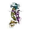
| ||||||||||||
|---|---|---|---|---|---|---|---|---|---|---|---|---|---|
| 1 |
| ||||||||||||
| 2 | 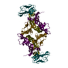
| ||||||||||||
| Unit cell |
| ||||||||||||
| Noncrystallographic symmetry (NCS) | NCS oper:
|
-Components
| #1: Protein | Mass: 11511.249 Da / Num. of mol.: 2 / Fragment: RECEPTOR BINDING DOMAIN, RESIDUES 8 - 109 Source method: isolated from a genetically manipulated source Source: (gene. exp.) Homo sapiens (human) / Production host:  #2: Protein | Mass: 10906.662 Da / Num. of mol.: 2 Fragment: SECOND EXTRACELLULAR IGG LIKE DOMAIN, RESIDUES 129 - 229 Source method: isolated from a genetically manipulated source Source: (gene. exp.) Homo sapiens (human) / Production host: References: UniProt: P17948, receptor protein-tyrosine kinase #3: Water | ChemComp-HOH / |  Mass: 18.015 Da / Num. of mol.: 476 / Source method: isolated from a natural source / Formula: H2O Mass: 18.015 Da / Num. of mol.: 476 / Source method: isolated from a natural source / Formula: H2OHas protein modification | Y | |
|---|
-Experimental details
-Experiment
| Experiment | Method: X-RAY DIFFRACTION / Number of used crystals: 1 |
|---|
- Sample preparation
| Crystal | Density Matthews: 2.3 Å3/Da / Density % sol: 47 % | ||||||||||||||||||||||||||||||||||||||||||
|---|---|---|---|---|---|---|---|---|---|---|---|---|---|---|---|---|---|---|---|---|---|---|---|---|---|---|---|---|---|---|---|---|---|---|---|---|---|---|---|---|---|---|---|
| Crystal grow | pH: 8.5 / Details: pH 8.5 | ||||||||||||||||||||||||||||||||||||||||||
| Crystal grow | *PLUS Method: vapor diffusion, hanging dropDetails: drop solution was mixed with an equal volume of reservoir solution | ||||||||||||||||||||||||||||||||||||||||||
| Components of the solutions | *PLUS
|
-Data collection
| Diffraction | Mean temperature: 160 K |
|---|---|
| Diffraction source | Source: SYNCHROTRON / Site: CHESS  / Beamline: A1 / Wavelength: 0.908 / Beamline: A1 / Wavelength: 0.908 |
| Detector | Type: ADSC QUANTUM / Detector: CCD / Date: Feb 1, 1997 |
| Radiation | Monochromatic (M) / Laue (L): M / Scattering type: x-ray |
| Radiation wavelength | Wavelength: 0.908 Å / Relative weight: 1 |
| Reflection | Resolution: 1.7→30 Å / Num. obs: 47047 / % possible obs: 99 % / Redundancy: 7 % / Biso Wilson estimate: 23.9 Å2 / Rsym value: 0.052 / Net I/σ(I): 8.3 |
| Reflection shell | Resolution: 1.7→1.76 Å / Redundancy: 3 % / Mean I/σ(I) obs: 3.5 / Rsym value: 0.27 / % possible all: 95.8 |
| Reflection | *PLUS Num. measured all: 332877 / Rmerge(I) obs: 0.052 |
| Reflection shell | *PLUS % possible obs: 95.8 % / Num. unique obs: 4520 / Rmerge(I) obs: 0.277 |
- Processing
| Software |
| ||||||||||||||||||||||||||||||||||||||||||||||||||||||||||||||||||||||||||||||||
|---|---|---|---|---|---|---|---|---|---|---|---|---|---|---|---|---|---|---|---|---|---|---|---|---|---|---|---|---|---|---|---|---|---|---|---|---|---|---|---|---|---|---|---|---|---|---|---|---|---|---|---|---|---|---|---|---|---|---|---|---|---|---|---|---|---|---|---|---|---|---|---|---|---|---|---|---|---|---|---|---|---|
| Refinement | Method to determine structure: MOLECULAR REPLACEMENT/NCS AVERAGING Starting model: 1VPF Resolution: 1.7→20 Å / Rfactor Rfree error: 0.005 / Data cutoff high absF: 1000000 / Data cutoff low absF: 0.001 / Isotropic thermal model: RESTRAINED / Cross valid method: THROUGHOUT / σ(F): 0.2
| ||||||||||||||||||||||||||||||||||||||||||||||||||||||||||||||||||||||||||||||||
| Displacement parameters | Biso mean: 31.9 Å2
| ||||||||||||||||||||||||||||||||||||||||||||||||||||||||||||||||||||||||||||||||
| Refine analyze |
| ||||||||||||||||||||||||||||||||||||||||||||||||||||||||||||||||||||||||||||||||
| Refinement step | Cycle: LAST / Resolution: 1.7→20 Å
| ||||||||||||||||||||||||||||||||||||||||||||||||||||||||||||||||||||||||||||||||
| Refine LS restraints |
| ||||||||||||||||||||||||||||||||||||||||||||||||||||||||||||||||||||||||||||||||
| LS refinement shell | Resolution: 1.7→1.78 Å / Rfactor Rfree error: 0.021 / Total num. of bins used: 8
| ||||||||||||||||||||||||||||||||||||||||||||||||||||||||||||||||||||||||||||||||
| Software | *PLUS Name: X-PLOR / Version: 3.851 / Classification: refinement | ||||||||||||||||||||||||||||||||||||||||||||||||||||||||||||||||||||||||||||||||
| Refinement | *PLUS | ||||||||||||||||||||||||||||||||||||||||||||||||||||||||||||||||||||||||||||||||
| Solvent computation | *PLUS | ||||||||||||||||||||||||||||||||||||||||||||||||||||||||||||||||||||||||||||||||
| Displacement parameters | *PLUS | ||||||||||||||||||||||||||||||||||||||||||||||||||||||||||||||||||||||||||||||||
| Refine LS restraints | *PLUS
| ||||||||||||||||||||||||||||||||||||||||||||||||||||||||||||||||||||||||||||||||
| LS refinement shell | *PLUS Rfactor obs: 0.32 |
