 Movie
Movie Controller


+ Open data

- Basic information
| Entry | Database: PDB / ID: 1vi5 | ||||||
|---|---|---|---|---|---|---|---|
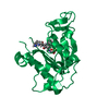
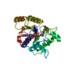
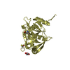
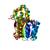
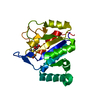
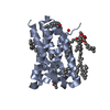
| Title | Crystal structure of ribosomal protein S2P | ||||||
 Components Components | 30S ribosomal protein S2P | ||||||
 Keywords Keywords | RIBOSOME / structural genomics | ||||||
| Function / homology |  Function and homology information Function and homology informationsmall ribosomal subunit / structural constituent of ribosome / translation Similarity search - Function | ||||||
| Biological species |   Archaeoglobus fulgidus (archaea) Archaeoglobus fulgidus (archaea) | ||||||
| Method |  X-RAY DIFFRACTION / SYNCHROTRON / Se-Met MAD phasing / Resolution: 2.65 Å X-RAY DIFFRACTION / SYNCHROTRON / Se-Met MAD phasing / Resolution: 2.65 Å | ||||||
 Authors Authors | Structural GenomiX | ||||||
 Citation Citation | Journal: Proteins / Year: 2005 Title: Structural analysis of a set of proteins resulting from a bacterial genomics project Authors: Badger, J. / Sauder, J.M. / Adams, J.M. / Antonysamy, S. / Bain, K. / Bergseid, M.G. / Buchanan, S.G. / Buchanan, M.D. / Batiyenko, Y. / Christopher, J.A. / Emtage, S. / Eroshkina, A. / ...Authors: Badger, J. / Sauder, J.M. / Adams, J.M. / Antonysamy, S. / Bain, K. / Bergseid, M.G. / Buchanan, S.G. / Buchanan, M.D. / Batiyenko, Y. / Christopher, J.A. / Emtage, S. / Eroshkina, A. / Feil, I. / Furlong, E.B. / Gajiwala, K.S. / Gao, X. / He, D. / Hendle, J. / Huber, A. / Hoda, K. / Kearins, P. / Kissinger, C. / Laubert, B. / Lewis, H.A. / Lin, J. / Loomis, K. / Lorimer, D. / Louie, G. / Maletic, M. / Marsh, C.D. / Miller, I. / Molinari, J. / Muller-Dieckmann, H.J. / Newman, J.M. / Noland, B.W. / Pagarigan, B. / Park, F. / Peat, T.S. / Post, K.W. / Radojicic, S. / Ramos, A. / Romero, R. / Rutter, M.E. / Sanderson, W.E. / Schwinn, K.D. / Tresser, J. / Winhoven, J. / Wright, T.A. / Wu, L. / Xu, J. / Harris, T.J. | ||||||
| History |
| ||||||
| Remark 300 | BIOMOLECULE: THIS ENTRY CONTAINS THE CRYSTALLOGRAPHIC ASYMMETRIC UNIT WHICH CONSISTS OF 4 CHAIN(S). ...BIOMOLECULE: THIS ENTRY CONTAINS THE CRYSTALLOGRAPHIC ASYMMETRIC UNIT WHICH CONSISTS OF 4 CHAIN(S). THE AUTHORS STATE THE BIOLOGICAL UNIT IS NOT KNOWN. |
- Structure visualization
| Structure viewer | Molecule: MolmilJmol/JSmol |
|---|
- Downloads & links
-Download
| PDBx/mmCIF format | 1vi5.cif.gz | 166.3 KB | Display | PDBx/mmCIF format |
|---|---|---|---|---|
| PDB format | pdb1vi5.ent.gz | 133 KB | Display | PDB format |
| PDBx/mmJSON format | 1vi5.json.gz | Tree view | PDBx/mmJSON format | |
| Others |  Other downloads Other downloads |
-Validation report
| Arichive directory | https://data.pdbj.org/pub/pdb/validation_reports/vi/1vi5ftp://data.pdbj.org/pub/pdb/validation_reports/vi/1vi5 | HTTPS FTP |
|---|
-Related structure data
| Related structure data | 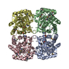 1o60C  1o61C  1o62C  1o63C  1o64C  1o65C  1o66C  1o67C 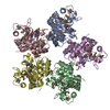 1o68C  1o69C  1o6bC  1o6cC  1o6dC  1vgtC  1vguC  1vgvC  1vgwC  1vgxC  1vgyC  1vgzC 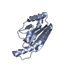 1vh0C  1vh1C  1vh2C  1vh3C  1vh4C  1vh5C 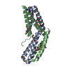 1vh6C  1vh7C  1vh8C  1vh9C  1vhaC  1vhcC  1vhdC 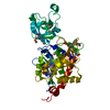 1vheC  1vhfC 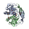 1vhgC  1vhjC  1vhkC  1vhlC  1vhmC  1vhoC 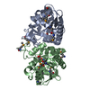 1vhqC  1vhsC 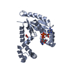 1vhtC  1vhuC 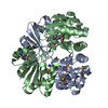 1vhvC  1vhwC  1vhxC 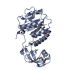 1vhyC 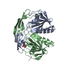 1vhzC 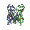 1vi0C  1vi1C  1vi2C 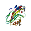 1vi3C 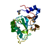 1vi4C  1vi6C  1vi8C 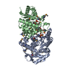 1vi9C  1viaC  1vicC  1vimC  1viqC  1visC  1viuC  1vivC  1vixC 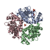 1viyC  1vizC C: citing same article ( |
|---|---|
| Similar structure data |

-Links
 PDBj
PDBj

- Assembly
| Deposited unit | 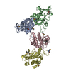
| ||||||||
|---|---|---|---|---|---|---|---|---|---|
| 1 | 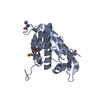
| ||||||||
| 2 | 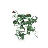
| ||||||||
| 3 | 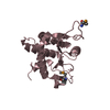
| ||||||||
| 4 | 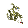
| ||||||||
| Unit cell |
|
-Components
| #1: Protein | Mass: 23663.439 Da / Num. of mol.: 4 Source method: isolated from a genetically manipulated source Source: (gene. exp.) Archaeoglobus fulgidus (archaea) / Gene: RPS2P, AF1133 / Production host:  #2: Water | ChemComp-HOH / |  Mass: 18.015 Da / Num. of mol.: 302 / Source method: isolated from a natural source / Formula: H2O Mass: 18.015 Da / Num. of mol.: 302 / Source method: isolated from a natural source / Formula: H2OHas protein modification | Y | |
|---|
-Experimental details
-Experiment
| Experiment | Method: X-RAY DIFFRACTION / Number of used crystals: 1 |
|---|
- Sample preparation
| Crystal | Density Matthews: 2.99 Å3/Da / Density % sol: 58.82 % | |||||||||||||||||||||||||||||||||||||||||||||||||
|---|---|---|---|---|---|---|---|---|---|---|---|---|---|---|---|---|---|---|---|---|---|---|---|---|---|---|---|---|---|---|---|---|---|---|---|---|---|---|---|---|---|---|---|---|---|---|---|---|---|---|
| Crystal grow | *PLUS pH: 7.5 / Method: vapor diffusion, hanging drop | |||||||||||||||||||||||||||||||||||||||||||||||||
| Components of the solutions | *PLUS
|
-Data collection
| Diffraction source | Source: SYNCHROTRON / Site: APS  / Beamline: 32-ID / Wavelength: 0.9795, 0.9641 / Beamline: 32-ID / Wavelength: 0.9795, 0.9641 | |||||||||
|---|---|---|---|---|---|---|---|---|---|---|
| Detector | Type: MARRESEARCH / Detector: CCD | |||||||||
| Radiation | Protocol: MAD / Scattering type: x-ray | |||||||||
| Radiation wavelength |
| |||||||||
| Reflection | Resolution: 2.65→54.7 Å / Num. all: 33692 / Num. obs: 33692 / % possible obs: 100 % / Redundancy: 6.6 % / Rmerge(I) obs: 0.131 / Net I/σ(I): 9.7 | |||||||||
| Reflection shell | Resolution: 2.65→2.79 Å / Redundancy: 6.6 % / Rmerge(I) obs: 0.736 / Mean I/σ(I) obs: 3.1 / % possible all: 100 | |||||||||
| Reflection shell | *PLUS % possible obs: 100 % |
- Processing
| Software |
| ||||||||||||||||||||||||||||||
|---|---|---|---|---|---|---|---|---|---|---|---|---|---|---|---|---|---|---|---|---|---|---|---|---|---|---|---|---|---|---|---|
| Refinement | Method to determine structure: Se-Met MAD phasing / Resolution: 2.65→54.7 Å / σ(F): 0
| ||||||||||||||||||||||||||||||
| Solvent computation | Solvent model: Babinet bulk solvent correction / Bsol: 490 Å2 / ksol: 0.966 e/Å3 | ||||||||||||||||||||||||||||||
| Displacement parameters | Biso mean: 41.612 Å2
| ||||||||||||||||||||||||||||||
| Refine Biso | Class: all / Treatment: isotropic | ||||||||||||||||||||||||||||||
| Refinement step | Cycle: LAST / Resolution: 2.65→54.7 Å
| ||||||||||||||||||||||||||||||
| Refine LS restraints |
| ||||||||||||||||||||||||||||||
| Software | *PLUS Version: 4 / Classification: refinement | ||||||||||||||||||||||||||||||
| Refine LS restraints | *PLUS
|
