 Movie
Movie Controller

[English] 日本語
 Yorodumi
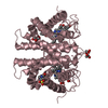
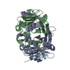
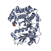
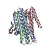
- PDB-3dcm: Crystal structure of the Thermotoga maritima SPOUT family RNA-met... -
+ Open data

- Basic information
| Entry | Database: PDB / ID: 3dcm | ||||||
|---|---|---|---|---|---|---|---|
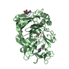
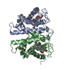
| Title | Crystal structure of the Thermotoga maritima SPOUT family RNA-methyltransferase protein Tm1570 in complex with S-adenosyl-L-methionine | ||||||
 Components Components | Uncharacterized protein TM_1570 | ||||||
 Keywords Keywords | TRANSFERASE / Trefoil knot / SPOUT Mtase / AdoMet binding | ||||||
| Function / homology |  Function and homology information Function and homology informationtRNA (guanine-N(1)-)-methyltransferase, C-terminal / SAM-dependent RNA methyltransferase / SPOUT methyltransferase, trefoil knot domain / Alpha/beta knot / tRNA (guanine-N1-)-methyltransferase, N-terminal / Alpha/beta knot methyltransferases / 3-Layer(aba) Sandwich / Alpha Beta Similarity search - Domain/homology | ||||||
| Biological species |   Thermotoga maritima (bacteria) Thermotoga maritima (bacteria) | ||||||
| Method |  X-RAY DIFFRACTION / SYNCHROTRON / MAD / Resolution: 2 Å X-RAY DIFFRACTION / SYNCHROTRON / MAD / Resolution: 2 Å | ||||||
 Authors Authors | Kim, D.J. / Kim, H.S. / Lee, S.J. / Suh, S.W. | ||||||
 Citation Citation | Journal: Proteins / Year: 2009 Title: Crystal structure of Thermotoga maritima SPOUT superfamily RNA methyltransferase Tm1570 in complex with S-adenosyl-L-methionine Authors: Kim, D.J. / Kim, H.S. / Lee, S.J. / Suh, S.W. | ||||||
| History |
|
- Structure visualization
| Structure viewer | Molecule: MolmilJmol/JSmol |
|---|
- Downloads & links
-Download
| PDBx/mmCIF format | 3dcm.cif.gz | 54.5 KB | Display | PDBx/mmCIF format |
|---|---|---|---|---|
| PDB format | pdb3dcm.ent.gz | 39 KB | Display | PDB format |
| PDBx/mmJSON format | 3dcm.json.gz | Tree view | PDBx/mmJSON format | |
| Others |  Other downloads Other downloads |
-Validation report
| Arichive directory | https://data.pdbj.org/pub/pdb/validation_reports/dc/3dcmftp://data.pdbj.org/pub/pdb/validation_reports/dc/3dcm | HTTPS FTP |
|---|
-Related structure data
| Similar structure data |
|---|


-Links
 PDBj
PDBj
- Assembly
| Deposited unit | 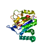
| ||||||||
|---|---|---|---|---|---|---|---|---|---|
| 1 | 
| ||||||||
| Unit cell |
| ||||||||
| Components on special symmetry positions |
|
-Components
| #1: Protein | Mass: 22008.250 Da / Num. of mol.: 1 Source method: isolated from a genetically manipulated source Source: (gene. exp.) Thermotoga maritima (bacteria) / Gene: TM_1570 / Plasmid: pET28b / Production host: |
|---|---|
| #2: Chemical | ChemComp-SAM /   Mass: 398.437 Da / Num. of mol.: 1 / Source method: obtained synthetically / Formula: C15H22N6O5S Mass: 398.437 Da / Num. of mol.: 1 / Source method: obtained synthetically / Formula: C15H22N6O5S |
| #3: Water | ChemComp-HOH /  Mass: 18.015 Da / Num. of mol.: 128 / Source method: isolated from a natural source / Formula: H2O Mass: 18.015 Da / Num. of mol.: 128 / Source method: isolated from a natural source / Formula: H2O |
-Experimental details
-Experiment
| Experiment | Method: X-RAY DIFFRACTION / Number of used crystals: 1 |
|---|
- Sample preparation
| Crystal | Density Matthews: 2.43 Å3/Da / Density % sol: 49.37 % / Mosaicity: 0.906 ° |
|---|---|
| Crystal grow | Temperature: 297 K / Method: vapor diffusion, hanging drop / pH: 7 Details: 100mM Tris-HCl pH 7.0, 200mM calcium acetate, 20% (v/v) PEG 3000, VAPOR DIFFUSION, HANGING DROP, temperature 297K |
-Data collection
| Diffraction |
| |||||||||||||||
|---|---|---|---|---|---|---|---|---|---|---|---|---|---|---|---|---|
| Diffraction source |
| |||||||||||||||
| Detector |
| |||||||||||||||
| Radiation |
| |||||||||||||||
| Radiation wavelength |
| |||||||||||||||
| Reflection | Resolution: 2→20 Å / Num. obs: 14038 / % possible obs: 98.4 % / Redundancy: 4.6 % / Rmerge(I) obs: 0.071 / Χ2: 2.005 / Net I/σ(I): 14.5 | |||||||||||||||
| Reflection shell | Resolution: 2→2.07 Å / Redundancy: 4.1 % / Rmerge(I) obs: 0.247 / Num. unique all: 1282 / Χ2: 1.432 / % possible all: 91.2 |

- Processing
| Software |
| ||||||||||||||||||||||||||||||||||||||||||||||||||||||||||||||||||||||||||||||||||||||||||
|---|---|---|---|---|---|---|---|---|---|---|---|---|---|---|---|---|---|---|---|---|---|---|---|---|---|---|---|---|---|---|---|---|---|---|---|---|---|---|---|---|---|---|---|---|---|---|---|---|---|---|---|---|---|---|---|---|---|---|---|---|---|---|---|---|---|---|---|---|---|---|---|---|---|---|---|---|---|---|---|---|---|---|---|---|---|---|---|---|---|---|---|
| Refinement | Method to determine structure: MAD / Resolution: 2→17.43 Å / Cor.coef. Fo:Fc: 0.942 / Cor.coef. Fo:Fc free: 0.93 / SU B: 3.885 / SU ML: 0.111 / Cross valid method: THROUGHOUT / σ(F): 0 / ESU R: 0.218 / ESU R Free: 0.178 / Stereochemistry target values: MAXIMUM LIKELIHOOD
| ||||||||||||||||||||||||||||||||||||||||||||||||||||||||||||||||||||||||||||||||||||||||||
| Solvent computation | Ion probe radii: 0.8 Å / Shrinkage radii: 0.8 Å / VDW probe radii: 1.4 Å / Solvent model: MASK | ||||||||||||||||||||||||||||||||||||||||||||||||||||||||||||||||||||||||||||||||||||||||||
| Displacement parameters | Biso mean: 24.379 Å2
| ||||||||||||||||||||||||||||||||||||||||||||||||||||||||||||||||||||||||||||||||||||||||||
| Refinement step | Cycle: LAST / Resolution: 2→17.43 Å
| ||||||||||||||||||||||||||||||||||||||||||||||||||||||||||||||||||||||||||||||||||||||||||
| Refine LS restraints |
| ||||||||||||||||||||||||||||||||||||||||||||||||||||||||||||||||||||||||||||||||||||||||||
| LS refinement shell | Resolution: 2.001→2.053 Å / Total num. of bins used: 20
|
