 Movie
Movie Controller

[English] 日本語
 Yorodumi
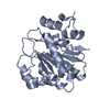
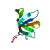
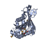
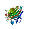
- PDB-2way: Structure of the human DDX6 C-terminal domain in complex with an ... -
+ Open data

- Basic information
| Entry | Database: PDB / ID: 2way | ||||||
|---|---|---|---|---|---|---|---|
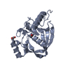
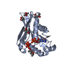
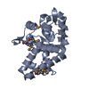
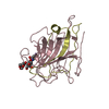
| Title | Structure of the human DDX6 C-terminal domain in complex with an EDC3- FDF peptide | ||||||
 Components Components |
| ||||||
 Keywords Keywords | HYDROLASE / DEAD-BOX PROTEIN / NUCLEOTIDE-BINDING / P54 / RCK / MIRNA / P-BODIES / HELICASE / DECAPPING / RNA-BINDING / PROTO-ONCOGENE / PHOSPHOPROTEIN / CHROMOSOMAL REARRANGEMENT / ATP-DEPENDENT RNA HELICASE / CYTOPLASM / MRNA DECAY / ATP-BINDING | ||||||
| Function / homology |  Function and homology information Function and homology informationconcave side of sperm head / outer dense fiber / deadenylation-independent decapping of nuclear-transcribed mRNA / spermatid differentiation / sperm annulus / mRNA decay by 5' to 3' exoribonuclease / chromatoid body / viral RNA genome packaging / miRNA-mediated gene silencing by inhibition of translation / RISC complex ...concave side of sperm head / outer dense fiber / deadenylation-independent decapping of nuclear-transcribed mRNA / spermatid differentiation / sperm annulus / mRNA decay by 5' to 3' exoribonuclease / chromatoid body / viral RNA genome packaging / miRNA-mediated gene silencing by inhibition of translation / RISC complex / P-body assembly / negative regulation of neuron differentiation / stem cell population maintenance / heterochromatin / stress granule assembly / helicase activity / adherens junction / P-body / neuron differentiation / cytoplasmic stress granule / cytoplasmic ribonucleoprotein granule / RNA helicase activity / negative regulation of translation / RNA helicase / cadherin binding / protein domain specific binding / mRNA binding / perinuclear region of cytoplasm / ATP hydrolysis activity / RNA binding / ATP binding / membrane / identical protein binding / nucleus / plasma membrane / cytoplasm / cytosol Similarity search - Function | ||||||
| Biological species |  HOMO SAPIENS (human) HOMO SAPIENS (human) | ||||||
| Method |  X-RAY DIFFRACTION / SYNCHROTRON / MOLECULAR REPLACEMENT / Resolution: 2.3 Å X-RAY DIFFRACTION / SYNCHROTRON / MOLECULAR REPLACEMENT / Resolution: 2.3 Å | ||||||
 Authors Authors | Tritschler, F. / Weichenrieder, O. | ||||||
 Citation Citation | Journal: Mol.Cell / Year: 2009 Title: Structural Basis for the Mutually Exclusive Anchoring of P Body Components Edc3 and Tral to the Dead Box Protein Ddx6/Me31B. Authors: Tritschler, F. / Braun, J.E. / Eulalio, A. / Truffault, V. / Izaurralde, E. / Weichenrieder, O. | ||||||
| History |
|
- Structure visualization
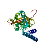
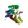
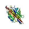
| Structure viewer | Molecule: MolmilJmol/JSmol |
|---|
- Downloads & links
-Download
| PDBx/mmCIF format | 2way.cif.gz | 90.2 KB | Display | PDBx/mmCIF format |
|---|---|---|---|---|
| PDB format | pdb2way.ent.gz | 68.1 KB | Display | PDB format |
| PDBx/mmJSON format | 2way.json.gz | Tree view | PDBx/mmJSON format | |
| Others |  Other downloads Other downloads |
-Validation report
| Arichive directory | https://data.pdbj.org/pub/pdb/validation_reports/wa/2wayftp://data.pdbj.org/pub/pdb/validation_reports/wa/2way | HTTPS FTP |
|---|
-Related structure data
| Related structure data | 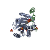 2waxC 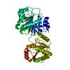 1s2mS C: citing same article ( S: Starting model for refinement |
|---|---|
| Similar structure data |

-Links
 PDBj
PDBj- Assembly
| Deposited unit | 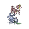
| ||||||||
|---|---|---|---|---|---|---|---|---|---|
| 1 | 
| ||||||||
| 2 | 
| ||||||||
| Unit cell |
|
-Components
| #1: Protein | Mass: 22475.650 Da / Num. of mol.: 2 / Fragment: C-TERMINAL DOMAIN, RESIDUES 296-483 Source method: isolated from a genetically manipulated source Source: (gene. exp.) HOMO SAPIENS (human) / Plasmid: PRSFDUET-1 / Production host:  References: UniProt: P26196, Hydrolases; Acting on acid anhydrides; In phosphorus-containing anhydrides #2: Protein/peptide | Mass: 5069.437 Da / Num. of mol.: 2 / Fragment: FDF PEPTIDE, RESIDUES 192-228 Source method: isolated from a genetically manipulated source Source: (gene. exp.) HOMO SAPIENS (human) / Plasmid: PRSFDUET-1 / Production host: #3: Chemical | ChemComp-GOL /   Mass: 92.094 Da / Num. of mol.: 4 / Source method: obtained synthetically / Formula: C3H8O3 Mass: 92.094 Da / Num. of mol.: 4 / Source method: obtained synthetically / Formula: C3H8O3#4: Water | ChemComp-HOH / |  Mass: 18.015 Da / Num. of mol.: 111 / Source method: isolated from a natural source / Formula: H2O Mass: 18.015 Da / Num. of mol.: 111 / Source method: isolated from a natural source / Formula: H2O |
|---|
-Experimental details
-Experiment
| Experiment | Method: X-RAY DIFFRACTION / Number of used crystals: 1 |
|---|
- Sample preparation
| Crystal | Density Matthews: 2.04 Å3/Da / Density % sol: 39.8 % / Description: NONE |
|---|---|
| Crystal grow | Details: 0.04 M KH2PO4, 16% PEG 8000, 20% GLYCEROL |
-Data collection
| Diffraction | Mean temperature: 90 K |
|---|---|
| Diffraction source | Source: SYNCHROTRON / Site: SLS  / Beamline: X10SA / Wavelength: 0.978 / Beamline: X10SA / Wavelength: 0.978 |
| Detector | Type: MARRESEARCH / Detector: CCD / Date: Jun 19, 2008 / Details: MIRRORS |
| Radiation | Monochromator: SI(111)MONOCHROMATOR / Protocol: SINGLE WAVELENGTH / Monochromatic (M) / Laue (L): M / Scattering type: x-ray |
| Radiation wavelength | Wavelength: 0.978 Å / Relative weight: 1 |
| Reflection | Resolution: 2.3→45.5 Å / Num. obs: 17392 / % possible obs: 95.5 % / Observed criterion σ(I): 0 / Redundancy: 4.6 % / Biso Wilson estimate: 38.6 Å2 / Rmerge(I) obs: 0.07 / Net I/σ(I): 16.5 |
| Reflection shell | Resolution: 2.3→2.36 Å / Redundancy: 4.5 % / Rmerge(I) obs: 0.36 / Mean I/σ(I) obs: 4.2 / % possible all: 98.2 |
- Processing
| Software |
| ||||||||||||||||||||||||||||||||||||||||||||||||||||||||||||||||||||||||||||||||||||||||||||||||||||||||||||||||||||||||||||||||||||||||||||||||||||||||||||||||||||||||||||||||||||||
|---|---|---|---|---|---|---|---|---|---|---|---|---|---|---|---|---|---|---|---|---|---|---|---|---|---|---|---|---|---|---|---|---|---|---|---|---|---|---|---|---|---|---|---|---|---|---|---|---|---|---|---|---|---|---|---|---|---|---|---|---|---|---|---|---|---|---|---|---|---|---|---|---|---|---|---|---|---|---|---|---|---|---|---|---|---|---|---|---|---|---|---|---|---|---|---|---|---|---|---|---|---|---|---|---|---|---|---|---|---|---|---|---|---|---|---|---|---|---|---|---|---|---|---|---|---|---|---|---|---|---|---|---|---|---|---|---|---|---|---|---|---|---|---|---|---|---|---|---|---|---|---|---|---|---|---|---|---|---|---|---|---|---|---|---|---|---|---|---|---|---|---|---|---|---|---|---|---|---|---|---|---|---|---|
| Refinement | Method to determine structure: MOLECULAR REPLACEMENT Starting model: PDB ENTRY 1S2M Resolution: 2.3→45.5 Å / Cor.coef. Fo:Fc: 0.927 / Cor.coef. Fo:Fc free: 0.875 / Cross valid method: THROUGHOUT / ESU R: 0.391 / ESU R Free: 0.275 / Stereochemistry target values: MAXIMUM LIKELIHOOD Details: HYDROGENS HAVE BEEN ADDED IN THE RIDING POSITIONS.RESIDUES A 383-387, A 413-419, A 461-472, B 192-198, B 225-228, C 296, C 383-389, C 413-419, C 463-472, D 192-195, D 226--228 ARE DISORDERED
| ||||||||||||||||||||||||||||||||||||||||||||||||||||||||||||||||||||||||||||||||||||||||||||||||||||||||||||||||||||||||||||||||||||||||||||||||||||||||||||||||||||||||||||||||||||||
| Solvent computation | Ion probe radii: 0.8 Å / Shrinkage radii: 0.8 Å / VDW probe radii: 1.2 Å / Solvent model: MASK | ||||||||||||||||||||||||||||||||||||||||||||||||||||||||||||||||||||||||||||||||||||||||||||||||||||||||||||||||||||||||||||||||||||||||||||||||||||||||||||||||||||||||||||||||||||||
| Displacement parameters | Biso mean: 33.48 Å2
| ||||||||||||||||||||||||||||||||||||||||||||||||||||||||||||||||||||||||||||||||||||||||||||||||||||||||||||||||||||||||||||||||||||||||||||||||||||||||||||||||||||||||||||||||||||||
| Refinement step | Cycle: LAST / Resolution: 2.3→45.5 Å
| ||||||||||||||||||||||||||||||||||||||||||||||||||||||||||||||||||||||||||||||||||||||||||||||||||||||||||||||||||||||||||||||||||||||||||||||||||||||||||||||||||||||||||||||||||||||
| Refine LS restraints |
|