 Movie
Movie Controller

[English] 日本語
 Yorodumi
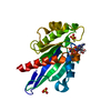
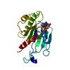
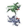
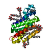
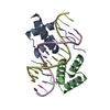
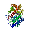
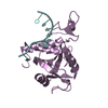
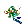
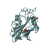
- PDB-6h35: Myxococcus xanthus MglA bound to GDP and Pi with mixed inactive a... -
+ Open data

- Basic information
| Entry | Database: PDB / ID: 6h35 | ||||||
|---|---|---|---|---|---|---|---|
| Title | Myxococcus xanthus MglA bound to GDP and Pi with mixed inactive and active switch region conformations | ||||||
 Components Components | Mutual gliding-motility protein MglA | ||||||
 Keywords Keywords | CYTOSOLIC PROTEIN / small GTPase in a mixed state | ||||||
| Function / homology |  Function and homology information Function and homology informationregulation of protein localization / GTPase activity / GTP binding / cytoplasm Similarity search - Function | ||||||
| Biological species |  Myxococcus xanthus DK 1622 (bacteria) Myxococcus xanthus DK 1622 (bacteria) | ||||||
| Method |  X-RAY DIFFRACTION / SYNCHROTRON / MOLECULAR REPLACEMENT / Resolution: 2.3 Å X-RAY DIFFRACTION / SYNCHROTRON / MOLECULAR REPLACEMENT / Resolution: 2.3 Å | ||||||
 Authors Authors | Varela, P.F. / Galicia, C. / Cherfils, J. | ||||||
| Funding support |  France, 1items France, 1items
| ||||||
 Citation Citation | Journal: Nat Commun / Year: 2019 Title: MglA functions as a three-state GTPase to control movement reversals of Myxococcus xanthus. Authors: Galicia, C. / Lhospice, S. / Varela, P.F. / Trapani, S. / Zhang, W. / Navaza, J. / Herrou, J. / Mignot, T. / Cherfils, J. | ||||||
| History |
|
- Structure visualization
| Structure viewer | Molecule: MolmilJmol/JSmol |
|---|
- Downloads & links
-Download
| PDBx/mmCIF format | 6h35.cif.gz | 211.8 KB | Display | PDBx/mmCIF format |
|---|---|---|---|---|
| PDB format | pdb6h35.ent.gz | 143 KB | Display | PDB format |
| PDBx/mmJSON format | 6h35.json.gz | Tree view | PDBx/mmJSON format | |
| Others |  Other downloads Other downloads |
-Validation report
| Arichive directory | https://data.pdbj.org/pub/pdb/validation_reports/h3/6h35ftp://data.pdbj.org/pub/pdb/validation_reports/h3/6h35 | HTTPS FTP |
|---|
-Related structure data


-Links
 PDBj
PDBj- Assembly
| Deposited unit | 
| ||||||||||||
|---|---|---|---|---|---|---|---|---|---|---|---|---|---|
| 1 | 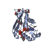
| ||||||||||||
| 2 | 
| ||||||||||||
| Unit cell |
| ||||||||||||
| Components on special symmetry positions |
|
-Components
| #1: Protein | Mass: 22871.262 Da / Num. of mol.: 2 Source method: isolated from a genetically manipulated source Details: Small GTPase / Source: (gene. exp.) Myxococcus xanthus DK 1622 (bacteria) / Gene: mglA, MXAN_1925 / Production host: #2: Chemical |   Type: RNA linking / Mass: 443.201 Da / Num. of mol.: 2 / Source method: obtained synthetically / Formula: C10H15N5O11P2 / Comment: GDP, energy-carrying molecule*YM Type: RNA linking / Mass: 443.201 Da / Num. of mol.: 2 / Source method: obtained synthetically / Formula: C10H15N5O11P2 / Comment: GDP, energy-carrying molecule*YM#3: Chemical | ChemComp-PO4 / |   Mass: 94.971 Da / Num. of mol.: 1 / Source method: obtained synthetically / Formula: PO4 Mass: 94.971 Da / Num. of mol.: 1 / Source method: obtained synthetically / Formula: PO4#4: Chemical | ChemComp-MPD / (   Mass: 118.174 Da / Num. of mol.: 9 / Source method: obtained synthetically / Formula: C6H14O2 / Comment: precipitant*YM Mass: 118.174 Da / Num. of mol.: 9 / Source method: obtained synthetically / Formula: C6H14O2 / Comment: precipitant*YM#5: Water | ChemComp-HOH / |  Mass: 18.015 Da / Num. of mol.: 155 / Source method: isolated from a natural source / Formula: H2O Mass: 18.015 Da / Num. of mol.: 155 / Source method: isolated from a natural source / Formula: H2O |
|---|
-Experimental details
-Experiment
| Experiment | Method: X-RAY DIFFRACTION / Number of used crystals: 1 |
|---|
- Sample preparation
| Crystal | Density Matthews: 2.77 Å3/Da / Density % sol: 55.66 % |
|---|---|
| Crystal grow | Temperature: 291 K / Method: vapor diffusion, hanging drop / pH: 7.5 / Details: 0.1 M HEPES pH 7.5, 70% MPD |
-Data collection
| Diffraction | Mean temperature: 100 K |
|---|---|
| Diffraction source | Source: SYNCHROTRON / Site: SOLEIL / Beamline: PROXIMA 1 / Wavelength: 0.98011 Å |
| Detector | Type: DECTRIS PILATUS 6M / Detector: PIXEL / Date: Jul 25, 2013 |
| Radiation | Protocol: SINGLE WAVELENGTH / Monochromatic (M) / Laue (L): M / Scattering type: x-ray |
| Radiation wavelength | Wavelength: 0.98011 Å / Relative weight: 1 |
| Reflection | Resolution: 2.299→50 Å / Num. obs: 93193 / % possible obs: 74.71 % / Redundancy: 5.3 % / Biso Wilson estimate: 38.07 Å2 / CC1/2: 0.99 / Rmerge(I) obs: 0.16 / Rpim(I) all: 0.076 / Net I/σ(I): 8.29 |
| Reflection shell | Resolution: 2.299→2.38 Å / Mean I/σ(I) obs: 1.03 / Num. unique obs: 474 / CC1/2: 0.392 / % possible all: 20.5 |
- Processing
| Software |
| |||||||||||||||||||||||||||||||||||||||||||||||||
|---|---|---|---|---|---|---|---|---|---|---|---|---|---|---|---|---|---|---|---|---|---|---|---|---|---|---|---|---|---|---|---|---|---|---|---|---|---|---|---|---|---|---|---|---|---|---|---|---|---|---|
| Refinement | Method to determine structure: MOLECULAR REPLACEMENT / Resolution: 2.3→49.11 Å / SU ML: 0.2609 / Cross valid method: FREE R-VALUE / σ(F): 1.34 / Phase error: 28.9156 / Stereochemistry target values: GeoStd + Monomer Library
| |||||||||||||||||||||||||||||||||||||||||||||||||
| Solvent computation | Shrinkage radii: 0.9 Å / VDW probe radii: 1.11 Å / Solvent model: FLAT BULK SOLVENT MODEL | |||||||||||||||||||||||||||||||||||||||||||||||||
| Displacement parameters | Biso mean: 44.8 Å2 | |||||||||||||||||||||||||||||||||||||||||||||||||
| Refinement step | Cycle: LAST / Resolution: 2.3→49.11 Å
| |||||||||||||||||||||||||||||||||||||||||||||||||
| Refine LS restraints |
| |||||||||||||||||||||||||||||||||||||||||||||||||
| LS refinement shell |
| |||||||||||||||||||||||||||||||||||||||||||||||||
| Refinement TLS params. | Method: refined / Origin x: 32.303813486 Å / Origin y: 14.7277344099 Å / Origin z: 70.125826273 Å
| |||||||||||||||||||||||||||||||||||||||||||||||||
| Refinement TLS group | Selection details: all |
