 Movie
Movie Controller

[English] 日本語
 Yorodumi
- PDB-2xow: Structure of GlpG in complex with a mechanism-based isocoumarin i... -
+ Open data

- Basic information
| Entry | Database: PDB / ID: 2xow | ||||||
|---|---|---|---|---|---|---|---|
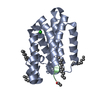
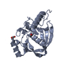
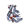
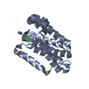
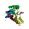
| Title | Structure of GlpG in complex with a mechanism-based isocoumarin inhibitor | ||||||
 Components Components | RHOMBOID PROTEASE GLPG | ||||||
 Keywords Keywords | HYDROLASE / MEMBRANE PROTEIN / INTRAMEMBRANE PROTEASE | ||||||
| Function / homology |  Function and homology information Function and homology informationrhomboid protease / endopeptidase activity / serine-type endopeptidase activity / proteolysis / identical protein binding / plasma membrane Similarity search - Function | ||||||
| Biological species |  | ||||||
| Method |  X-RAY DIFFRACTION / SYNCHROTRON / MOLECULAR REPLACEMENT / Resolution: 2.09 Å X-RAY DIFFRACTION / SYNCHROTRON / MOLECULAR REPLACEMENT / Resolution: 2.09 Å | ||||||
 Authors Authors | Vinothkumar, K.R. / Strisovsky, K. / Andreeva, A. / Christova, Y. / Verhelst, S. / Freeman, M. | ||||||
 Citation Citation | Journal: Embo J. / Year: 2010 Title: The Structural Basis for Catalysis and Substrate Specificity of a Rhomboid Protease Authors: Vinothkumar, K.R. / Strisovsky, K. / Andreeva, A. / Christova, Y. / Verhelst, S. / Freeman, M. | ||||||
| History |
|
- Structure visualization
| Structure viewer | Molecule: MolmilJmol/JSmol |
|---|
- Downloads & links
-Download
| PDBx/mmCIF format | 2xow.cif.gz | 55.2 KB | Display | PDBx/mmCIF format |
|---|---|---|---|---|
| PDB format | pdb2xow.ent.gz | 38.9 KB | Display | PDB format |
| PDBx/mmJSON format | 2xow.json.gz | Tree view | PDBx/mmJSON format | |
| Others |  Other downloads Other downloads |
-Validation report
| Arichive directory | https://data.pdbj.org/pub/pdb/validation_reports/xo/2xowftp://data.pdbj.org/pub/pdb/validation_reports/xo/2xow | HTTPS FTP |
|---|
-Related structure data
| Related structure data |  2xovC  3b45S C: citing same article ( S: Starting model for refinement |
|---|---|
| Similar structure data |


-Links
 PDBj
PDBj
- Assembly
| Deposited unit | 
| ||||||||
|---|---|---|---|---|---|---|---|---|---|
| 1 |
| ||||||||
| Unit cell |
|
-Components
| #1: Protein | Mass: 20214.020 Da / Num. of mol.: 1 / Fragment: CORE TM DOMAIN, RESIDUES 92-270 Source method: isolated from a genetically manipulated source Source: (gene. exp.) | ||||||
|---|---|---|---|---|---|---|---|
| #2: Chemical | ChemComp-ISM /   Mass: 209.199 Da / Num. of mol.: 1 / Source method: obtained synthetically / Formula: C10H11NO4 Mass: 209.199 Da / Num. of mol.: 1 / Source method: obtained synthetically / Formula: C10H11NO4 | ||||||
| #3: Sugar | ChemComp-BNG / 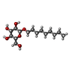  Type: D-saccharide / Mass: 306.395 Da / Num. of mol.: 16 Type: D-saccharide / Mass: 306.395 Da / Num. of mol.: 16Source method: isolated from a genetically manipulated source Formula: C15H30O6 / Comment: detergent*YM #4: Water | ChemComp-HOH / |  Mass: 18.015 Da / Num. of mol.: 38 / Source method: isolated from a natural source / Formula: H2O Mass: 18.015 Da / Num. of mol.: 38 / Source method: isolated from a natural source / Formula: H2OHas protein modification | Y | Nonpolymer details | METHYL-2(2-CARBOXY-4-AMINO-PHENYL)ACETATE (ISM): LINKS C1 OF ISM AND OG OF S201 AND C8 OF ISM AND ...METHYL-2(2-CARBOXY-4-AMINO-PHENYL)ACETATE (ISM): LINKS C1 OF ISM AND OG OF S201 AND C8 OF ISM AND NE2 OF H254 THE ISOCOUMARI | |
-Experimental details
-Experiment
| Experiment | Method: X-RAY DIFFRACTION / Number of used crystals: 1 |
|---|
- Sample preparation
| Crystal | Density Matthews: 3.5 Å3/Da / Density % sol: 63.47 % / Description: NONE |
|---|---|
| Crystal grow | Temperature: 298 K / Method: vapor diffusion, hanging drop / pH: 7 / Details: 2 M AMMONIUM CHLORIDE, 0.1 M BIS-TRIS PH 7.0, 298K |
-Data collection
| Diffraction | Mean temperature: 100 K |
|---|---|
| Diffraction source | Source: SYNCHROTRON / Site: Diamond  / Beamline: I02 / Wavelength: 0.9795 / Beamline: I02 / Wavelength: 0.9795 |
| Detector | Type: ADSC CCD / Detector: CCD / Date: Oct 11, 2009 |
| Radiation | Protocol: SINGLE WAVELENGTH / Monochromatic (M) / Laue (L): M / Scattering type: x-ray |
| Radiation wavelength | Wavelength: 0.9795 Å / Relative weight: 1 |
| Reflection | Resolution: 2.09→44.62 Å / Num. obs: 16662 / % possible obs: 97 % / Redundancy: 4.9 % / Biso Wilson estimate: 37.61 Å2 / Rmerge(I) obs: 0.05 / Net I/σ(I): 16.3 |
| Reflection shell | Resolution: 2.09→2.2 Å / Redundancy: 3.5 % / Rmerge(I) obs: 0.39 / Mean I/σ(I) obs: 2.9 / % possible all: 85.4 |
- Processing
| Software |
| |||||||||||||||||||||||||||||||||||||||||||||||||
|---|---|---|---|---|---|---|---|---|---|---|---|---|---|---|---|---|---|---|---|---|---|---|---|---|---|---|---|---|---|---|---|---|---|---|---|---|---|---|---|---|---|---|---|---|---|---|---|---|---|---|
| Refinement | Method to determine structure: MOLECULAR REPLACEMENT Starting model: PDB ENTRY 3B45 Resolution: 2.09→31.161 Å / SU ML: 0.24 / σ(F): 1.34 / Phase error: 23.42 / Stereochemistry target values: ML Details: COVALENT LINK OF ICM WITH OG OF SER 201 AND NE2 OF HIS 254
| |||||||||||||||||||||||||||||||||||||||||||||||||
| Solvent computation | Shrinkage radii: 0.9 Å / VDW probe radii: 1.11 Å / Solvent model: FLAT BULK SOLVENT MODEL / Bsol: 80.419 Å2 / ksol: 0.4 e/Å3 | |||||||||||||||||||||||||||||||||||||||||||||||||
| Displacement parameters | Biso mean: 44.72 Å2
| |||||||||||||||||||||||||||||||||||||||||||||||||
| Refinement step | Cycle: LAST / Resolution: 2.09→31.161 Å
| |||||||||||||||||||||||||||||||||||||||||||||||||
| Refine LS restraints |
| |||||||||||||||||||||||||||||||||||||||||||||||||
| LS refinement shell |
|
