 Movie
Movie Controller

[English] 日本語
 Yorodumi
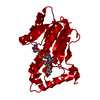
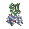
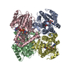
- PDB-1upf: STRUCTURE OF THE URACIL PHOSPHORIBOSYLTRANSFERASE, MUTANT C128V B... -
+ Open data

- Basic information
| Entry | Database: PDB / ID: 1upf | ||||||
|---|---|---|---|---|---|---|---|
| Title | STRUCTURE OF THE URACIL PHOSPHORIBOSYLTRANSFERASE, MUTANT C128V BOUND TO THE DRUG 5-FLUOROURACIL | ||||||
 Components Components | URACIL PHOSPHORIBOSYLTRANSFERASE | ||||||
 Keywords Keywords | TRANSFERASE / GLYCOSYLTRANSFERASE / PHOSPHORIBOSYLTRANSFERASE | ||||||
| Function / homology |  Function and homology information Function and homology informationuracil phosphoribosyltransferase / uracil phosphoribosyltransferase activity / UMP salvage / GTP binding Similarity search - Function | ||||||
| Biological species |  | ||||||
| Method |  X-RAY DIFFRACTION / Resolution: 2.3 Å X-RAY DIFFRACTION / Resolution: 2.3 Å | ||||||
 Authors Authors | Schumacher, M.A. / Carter, D. / Scott, D. / Roos, D. / Ullman, B. / Brennan, R.G. | ||||||
 Citation Citation | Journal: EMBO J. / Year: 1998 Title: Crystal structures of Toxoplasma gondii uracil phosphoribosyltransferase reveal the atomic basis of pyrimidine discrimination and prodrug binding. Authors: Schumacher, M.A. / Carter, D. / Scott, D.M. / Roos, D.S. / Ullman, B. / Brennan, R.G. | ||||||
| History |
|
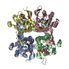
- Structure visualization
| Structure viewer | Molecule: MolmilJmol/JSmol |
|---|
- Downloads & links
-Download
| PDBx/mmCIF format | 1upf.cif.gz | 191.7 KB | Display | PDBx/mmCIF format |
|---|---|---|---|---|
| PDB format | pdb1upf.ent.gz | 154.4 KB | Display | PDB format |
| PDBx/mmJSON format | 1upf.json.gz | Tree view | PDBx/mmJSON format | |
| Others |  Other downloads Other downloads |
-Validation report
| Arichive directory | https://data.pdbj.org/pub/pdb/validation_reports/up/1upfftp://data.pdbj.org/pub/pdb/validation_reports/up/1upf | HTTPS FTP |
|---|
-Related structure data


-Links
 PDBj
PDBj
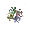
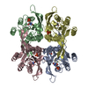
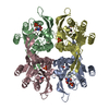
- Assembly
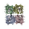
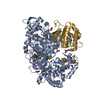
| Deposited unit | 
| ||||||||
|---|---|---|---|---|---|---|---|---|---|
| 1 |
| ||||||||
| Unit cell |
|
-Components
| #1: Protein | Mass: 25543.012 Da / Num. of mol.: 4 / Mutation: C128V Source method: isolated from a genetically manipulated source Source: (gene. exp.)  References: UniProt: Q26998, uracil phosphoribosyltransferase #2: Chemical | ChemComp-SO4 /   Mass: 96.063 Da / Num. of mol.: 12 / Source method: obtained synthetically / Formula: SO4 Mass: 96.063 Da / Num. of mol.: 12 / Source method: obtained synthetically / Formula: SO4#3: Chemical | ChemComp-URF / 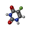  Mass: 130.077 Da / Num. of mol.: 4 / Source method: obtained synthetically / Formula: C4H3FN2O2 / Comment: medication, chemotherapy*YM Mass: 130.077 Da / Num. of mol.: 4 / Source method: obtained synthetically / Formula: C4H3FN2O2 / Comment: medication, chemotherapy*YM#4: Water | ChemComp-HOH / |  Mass: 18.015 Da / Num. of mol.: 207 / Source method: isolated from a natural source / Formula: H2O Mass: 18.015 Da / Num. of mol.: 207 / Source method: isolated from a natural source / Formula: H2ONonpolymer details | THE STRUCTURE HAS THREE SULPHATE MOLECULES (USED IN THE CRYSTALLIZATION). ONE SULPHATE IS BOUND IN ...THE STRUCTURE HAS THREE SULPHATE MOLECULES (USED IN THE CRYSTALLIZ | |
|---|
-Experimental details
-Experiment
| Experiment | Method: X-RAY DIFFRACTION |
|---|
- Sample preparation
| Crystal | Density Matthews: 2.72 Å3/Da / Density % sol: 54.7 % | ||||||||||||||||||||||||||||||||||||||||||
|---|---|---|---|---|---|---|---|---|---|---|---|---|---|---|---|---|---|---|---|---|---|---|---|---|---|---|---|---|---|---|---|---|---|---|---|---|---|---|---|---|---|---|---|
| Crystal | *PLUS Density % sol: 49 % | ||||||||||||||||||||||||||||||||||||||||||
| Crystal grow | *PLUS pH: 7.6 / Method: vapor diffusion, hanging drop | ||||||||||||||||||||||||||||||||||||||||||
| Components of the solutions | *PLUS
|
-Data collection
| Diffraction | Mean temperature: 298 K |
|---|---|
| Detector | Date: Jan 1, 1997 |
| Radiation | Monochromatic (M) / Laue (L): M / Scattering type: x-ray |
| Radiation wavelength | Relative weight: 1 |
| Reflection | Highest resolution: 2.3 Å |
| Reflection | *PLUS Lowest resolution: 10 Å / Num. obs: 62830 / % possible obs: 86 % / Num. measured all: 91812 / Rmerge(I) obs: 0.052 |
- Processing
| Software | Name: TNT / Classification: refinement | ||||||||||||||||||||||||||||||
|---|---|---|---|---|---|---|---|---|---|---|---|---|---|---|---|---|---|---|---|---|---|---|---|---|---|---|---|---|---|---|---|
| Refinement | Resolution: 2.3→10 Å / σ(F): 1 Details: THERE IS A TETRAMER (CHAINS A,B,C,D) IN THE ASYMMETRIC UNIT. THE P21 SPACE GROUP CAN BE TRANSFORMED INTO C2221. WITH TWO IN THE ASU. THE STRUCTURE WAS SOLVED AS P21 FOR TECHNICAL REASONS.
| ||||||||||||||||||||||||||||||
| Refinement step | Cycle: LAST / Resolution: 2.3→10 Å
| ||||||||||||||||||||||||||||||
| Refine LS restraints |
|
