 Movie
Movie Controller


+ Open data

- Basic information
| Entry | Database: PDB / ID: 1bd4 | ||||||
|---|---|---|---|---|---|---|---|
| Title | UPRT-URACIL COMPLEX | ||||||
 Components Components | URACIL PHOSPHORIBOSYLTRANSFERASE | ||||||
 Keywords Keywords | TRANSFERASE / UPRT / URACIL / GLYCOSYLTRANSFERASE / COMPLEX | ||||||
| Function / homology |  Function and homology information Function and homology informationuracil phosphoribosyltransferase / uracil phosphoribosyltransferase activity / UMP salvage / GTP binding Similarity search - Function | ||||||
| Biological species |  | ||||||
| Method |  X-RAY DIFFRACTION / Resolution: 2.2 Å X-RAY DIFFRACTION / Resolution: 2.2 Å | ||||||
 Authors Authors | Schumacher, M.A. / Carter, D. / Scott, D. / Roos, D. / Ullman, B. / Brennan, R.G. | ||||||
 Citation Citation | Journal: EMBO J. / Year: 1998 Title: Crystal structures of Toxoplasma gondii uracil phosphoribosyltransferase reveal the atomic basis of pyrimidine discrimination and prodrug binding. Authors: Schumacher, M.A. / Carter, D. / Scott, D.M. / Roos, D.S. / Ullman, B. / Brennan, R.G. | ||||||
| History |
|
- Structure visualization
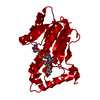
| Structure viewer | Molecule: MolmilJmol/JSmol |
|---|
- Downloads & links
-Download
| PDBx/mmCIF format | 1bd4.cif.gz | 191.6 KB | Display | PDBx/mmCIF format |
|---|---|---|---|---|
| PDB format | pdb1bd4.ent.gz | 153.8 KB | Display | PDB format |
| PDBx/mmJSON format | 1bd4.json.gz | Tree view | PDBx/mmJSON format | |
| Others |  Other downloads Other downloads |
-Validation report
| Arichive directory | https://data.pdbj.org/pub/pdb/validation_reports/bd/1bd4ftp://data.pdbj.org/pub/pdb/validation_reports/bd/1bd4 | HTTPS FTP |
|---|
-Related structure data


-Links
 PDBj
PDBj
- Assembly
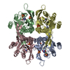
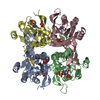
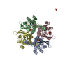
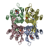
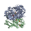
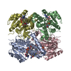
| Deposited unit | 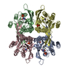
| ||||||||
|---|---|---|---|---|---|---|---|---|---|
| 1 |
| ||||||||
| Unit cell |
|
-Components
| #1: Protein | Mass: 27558.246 Da / Num. of mol.: 4 / Mutation: C128V Source method: isolated from a genetically manipulated source Source: (gene. exp.)  References: UniProt: Q26998, uracil phosphoribosyltransferase #2: Chemical | ChemComp-PO4 /   Mass: 94.971 Da / Num. of mol.: 8 / Source method: obtained synthetically / Formula: PO4 Mass: 94.971 Da / Num. of mol.: 8 / Source method: obtained synthetically / Formula: PO4#3: Chemical | ChemComp-URA /   Mass: 112.087 Da / Num. of mol.: 4 / Source method: obtained synthetically / Formula: C4H4N2O2 Mass: 112.087 Da / Num. of mol.: 4 / Source method: obtained synthetically / Formula: C4H4N2O2#4: Water | ChemComp-HOH / |  Mass: 18.015 Da / Num. of mol.: 240 / Source method: isolated from a natural source / Formula: H2O Mass: 18.015 Da / Num. of mol.: 240 / Source method: isolated from a natural source / Formula: H2ONonpolymer details | THE STRUCTURE HAS TWO PHOSPHATE MOLECULES (USED IN CRYSTALLIZATION). ONE PHOSPHATE FOUND IN THE ...THE STRUCTURE HAS TWO PHOSPHATE MOLECULES (USED IN CRYSTALLIZ | |
|---|
-Experimental details
-Experiment
| Experiment | Method: X-RAY DIFFRACTION |
|---|
- Sample preparation
| Crystal | Density Matthews: 2.52 Å3/Da / Density % sol: 51.14 % | ||||||||||||||||||||||||||||||||||||||||||
|---|---|---|---|---|---|---|---|---|---|---|---|---|---|---|---|---|---|---|---|---|---|---|---|---|---|---|---|---|---|---|---|---|---|---|---|---|---|---|---|---|---|---|---|
| Crystal | *PLUS Density % sol: 49 % | ||||||||||||||||||||||||||||||||||||||||||
| Crystal grow | *PLUS pH: 7.6 / Method: vapor diffusion, hanging drop | ||||||||||||||||||||||||||||||||||||||||||
| Components of the solutions | *PLUS
|
-Data collection
| Diffraction | Mean temperature: 298 K |
|---|---|
| Detector | Date: Jan 1, 1997 |
| Radiation | Monochromatic (M) / Laue (L): M / Scattering type: x-ray |
| Radiation wavelength | Relative weight: 1 |
| Reflection | Highest resolution: 2.2 Å |
| Reflection | *PLUS Highest resolution: 2.2 Å / Lowest resolution: 10 Å / Num. obs: 53303 / % possible obs: 87 % / Num. measured all: 111427 / Rmerge(I) obs: 0.052 |
| Reflection shell | *PLUS Highest resolution: 2.16 Å / Lowest resolution: 2.23 Å / Mean I/σ(I) obs: 3.9 |
- Processing
| Software | Name: TNT / Classification: refinement | ||||||||||||||||||||||||||||||
|---|---|---|---|---|---|---|---|---|---|---|---|---|---|---|---|---|---|---|---|---|---|---|---|---|---|---|---|---|---|---|---|
| Refinement | Resolution: 2.2→10 Å / σ(F): 1 Details: THERE IS A TETRAMER (CHAINS A, B, C, D) IN THE ASYMMETRIC UNIT. THERE ARE FOUR IN THE ASYMMETRIC UNIT FOR SPACE GROUP P21. P21 SPACE GROUP CAN BE TRANSFORMED TO C2221. SOLVED AS P21 FOR ...Details: THERE IS A TETRAMER (CHAINS A, B, C, D) IN THE ASYMMETRIC UNIT. THERE ARE FOUR IN THE ASYMMETRIC UNIT FOR SPACE GROUP P21. P21 SPACE GROUP CAN BE TRANSFORMED TO C2221. SOLVED AS P21 FOR TECHNICAL REASONS. THE N-TERMINAL MET IS CLEAVED OFF IN ESCHERICHIA COLI. RESIDUES 2 THROUGH 20 ARE DISORDERED AND NOT INCLUDED. STRUCTURE INCLUDES RESIDUES 21 TO 244 OF EACH MONOMER. THERE IS A TETRAMER (CHAINS A, B, C, D) IN THE ASYMMETRIC UNIT. THERE ARE FOUR IN THE ASYMMETRIC UNIT FOR SPACE GROUP P21. P21 SPACE GROUP CAN BE TRANSFORMED TO C2221. SOLVED AS P21 FOR TECHNICAL REASONS.
| ||||||||||||||||||||||||||||||
| Refinement step | Cycle: LAST / Resolution: 2.2→10 Å
| ||||||||||||||||||||||||||||||
| Refine LS restraints |
| ||||||||||||||||||||||||||||||
| Refinement | *PLUS Rfactor obs: 0.181 | ||||||||||||||||||||||||||||||
| Solvent computation | *PLUS | ||||||||||||||||||||||||||||||
| Displacement parameters | *PLUS |
