[English] 日本語
 Yorodumi
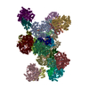
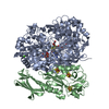
- PDB-4v4e: Crystal Structure of Pyrogallol-Phloroglucinol Transhydroxylase f... -
+ Open data
- Basic information
| Entry | Database: PDB / ID: 4v4e | |||||||||
|---|---|---|---|---|---|---|---|---|---|---|
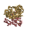
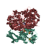
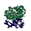
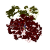
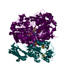
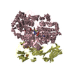
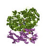
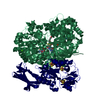
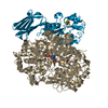
| Title | Crystal Structure of Pyrogallol-Phloroglucinol Transhydroxylase from Pelobacter acidigallici complexed with inhibitor 1,2,4,5-tetrahydroxy-benzene | |||||||||
 Components Components | (Pyrogallol hydroxytransferase ...) x 2 | |||||||||
 Keywords Keywords | OXIDOREDUCTASE / Molybdenum binding enzyme / MGD-cofactors / DMSO-reductase family / 4Fe-4S-cluster / complex with inhibitor 1 / 2 / 4 / 5-tetrahydroxy benzene | |||||||||
| Function / homology |  Function and homology information Function and homology informationpyrogallol hydroxytransferase / pyrogallol hydroxytransferase activity / molybdenum ion binding / molybdopterin cofactor binding / anaerobic respiration / outer membrane-bounded periplasmic space / 4 iron, 4 sulfur cluster binding / electron transfer activity / metal ion binding Similarity search - Function | |||||||||
| Biological species |  Pelobacter acidigallici (bacteria) Pelobacter acidigallici (bacteria) | |||||||||
| Method |  X-RAY DIFFRACTION / X-RAY DIFFRACTION /  SYNCHROTRON / SYNCHROTRON /  FOURIER SYNTHESIS / Resolution: 2 Å FOURIER SYNTHESIS / Resolution: 2 Å | |||||||||
 Authors Authors | Messerschmidt, A. / Niessen, H. / Abt, D. / Einsle, O. / Schink, B. / Kroneck, P.M.H. | |||||||||
 Citation Citation |  Journal: PROC.NATL.ACAD.SCI.USA / Year: 2004 Journal: PROC.NATL.ACAD.SCI.USA / Year: 2004Title: Crystal structure of pyrogallol-phloroglucinol transhydroxylase, an Mo enzyme capable of intermolecular hydroxyl transfer between phenols Authors: Messerschmidt, A. / Niessen, H. / Abt, D. / Einsle, O. / Schink, B. / Kroneck, P.M.H. | |||||||||
| History |
| |||||||||
| Remark 400 | THE COMPLETE CRYSTAL STRUCTURE IS REPRESENTED BY PDB STRUCTURES 1TI6 AND 1VLF |
- Structure visualization
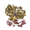
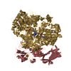
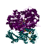
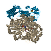
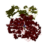
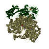
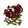
| Structure viewer | Molecule:  Molmil Molmil Jmol/JSmol Jmol/JSmol |
|---|
- Downloads & links
- Download
| PDBx/mmCIF format |  4v4e.cif.gz 4v4e.cif.gz | 2.9 MB | Display |  PDBx/mmCIF format PDBx/mmCIF format |
|---|---|---|---|---|
| PDB format |  pdb4v4e.ent.gz pdb4v4e.ent.gz | Display |  PDB format PDB format | |
| PDBx/mmJSON format |  4v4e.json.gz 4v4e.json.gz | Tree view |  PDBx/mmJSON format PDBx/mmJSON format | |
| Others |  Other downloads Other downloads |
-Validation report
| Arichive directory |  https://data.pdbj.org/pub/pdb/validation_reports/v4/4v4e https://data.pdbj.org/pub/pdb/validation_reports/v4/4v4e ftp://data.pdbj.org/pub/pdb/validation_reports/v4/4v4e ftp://data.pdbj.org/pub/pdb/validation_reports/v4/4v4e | HTTPS FTP |
|---|
-Related structure data
| Related structure data | 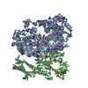 4v4cC 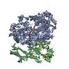 4v4dC  1ti2 C: citing same article ( S: Starting model for refinement |
|---|---|
| Similar structure data |
- Links
Links
- Assembly
- Components
-Pyrogallol hydroxytransferase ... , 2 types, 24 molecules ACEGIKMOQSUWBDFHJLNPRTVX
| #1: Protein | Mass: 99379.898 Da / Num. of mol.: 12 / Source method: isolated from a natural source / Source: (natural)  Pelobacter acidigallici (bacteria) / Strain: MaGal 2(DSM 2377) / References: UniProt: P80563, pyrogallol hydroxytransferase Pelobacter acidigallici (bacteria) / Strain: MaGal 2(DSM 2377) / References: UniProt: P80563, pyrogallol hydroxytransferase#2: Protein | Mass: 31259.424 Da / Num. of mol.: 12 / Source method: isolated from a natural source / Source: (natural)  Pelobacter acidigallici (bacteria) / Strain: MaGal 2(DSM 2377) / References: UniProt: P80564, pyrogallol hydroxytransferase Pelobacter acidigallici (bacteria) / Strain: MaGal 2(DSM 2377) / References: UniProt: P80564, pyrogallol hydroxytransferase |
|---|
-Non-polymers , 6 types, 10199 molecules 
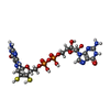

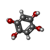


| #3: Chemical | ChemComp-CA / #4: Chemical | ChemComp-MGD / #5: Chemical | ChemComp-4MO / #6: Chemical | ChemComp-BTT / #7: Chemical | ChemComp-SF4 / #8: Water | ChemComp-HOH / | |
|---|
-Experimental details
-Experiment
| Experiment | Method:  X-RAY DIFFRACTION / Number of used crystals: 1 X-RAY DIFFRACTION / Number of used crystals: 1 |
|---|
- Sample preparation
| Crystal | Density Matthews: 2.71 Å3/Da / Density % sol: 55 % |
|---|---|
| Crystal grow | Temperature: 291 K / Method: vapor diffusion, sitting drop / pH: 7.5 Details: PEG 8000, sodium acetate, potassium phosphate, sodium dithionite, sodium cacodylate, pH 7.5, VAPOR DIFFUSION, SITTING DROP, temperature 291.0K |
-Data collection
| Diffraction | Mean temperature: 100 K |
|---|---|
| Diffraction source | Source:  SYNCHROTRON / Site: SYNCHROTRON / Site:  ESRF ESRF  / Beamline: ID29 / Wavelength: 0.9393 Å / Beamline: ID29 / Wavelength: 0.9393 Å |
| Detector | Type: ADSC QUANTUM 4 / Detector: CCD / Date: Dec 17, 2002 / Details: mirror |
| Radiation | Protocol: SINGLE WAVELENGTH / Monochromatic (M) / Laue (L): M / Scattering type: x-ray |
| Radiation wavelength | Wavelength: 0.9393 Å / Relative weight: 1 |
| Reflection | Resolution: 2→25 Å / Num. all: 1122947 / Num. obs: 1084767 / % possible obs: 96.6 % / Observed criterion σ(F): 2 / Observed criterion σ(I): 2 / Redundancy: 3.2 % / Biso Wilson estimate: 17.4 Å2 / Rmerge(I) obs: 0.12 / Rsym value: 0.12 / Net I/σ(I): 4 |
| Reflection shell | Resolution: 2→2.11 Å / Redundancy: 2.7 % / Rmerge(I) obs: 0.34 / Mean I/σ(I) obs: 1.9 / Rsym value: 0.34 / % possible all: 92 |
- Processing
| Software |
| ||||||||||||||||||||||||||||||||||||
|---|---|---|---|---|---|---|---|---|---|---|---|---|---|---|---|---|---|---|---|---|---|---|---|---|---|---|---|---|---|---|---|---|---|---|---|---|---|
| Refinement | Method to determine structure:  FOURIER SYNTHESIS FOURIER SYNTHESISStarting model: 1TI2  1ti2 Resolution: 2→24.99 Å / Rfactor Rfree error: 0.001 / Data cutoff high absF: 5564441.04 / Data cutoff low absF: 0 / Isotropic thermal model: RESTRAINED / Cross valid method: THROUGHOUT / σ(F): 0 / Stereochemistry target values: Engh & Huber
| ||||||||||||||||||||||||||||||||||||
| Solvent computation | Solvent model: FLAT MODEL / Bsol: 42.7191 Å2 / ksol: 0.358571 e/Å3 | ||||||||||||||||||||||||||||||||||||
| Displacement parameters | Biso mean: 19 Å2
| ||||||||||||||||||||||||||||||||||||
| Refine analyze |
| ||||||||||||||||||||||||||||||||||||
| Refinement step | Cycle: LAST / Resolution: 2→24.99 Å
| ||||||||||||||||||||||||||||||||||||
| Refine LS restraints |
| ||||||||||||||||||||||||||||||||||||
| LS refinement shell | Resolution: 2→2.13 Å / Rfactor Rfree error: 0.002 / Total num. of bins used: 6
| ||||||||||||||||||||||||||||||||||||
| Xplor file |
|
 Movie
Movie Controller








 PDBj
PDBj


