[English] 日本語
Yorodumi
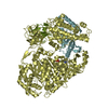
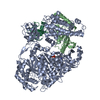
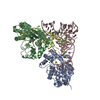
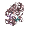
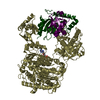
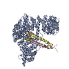
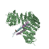
- PDB-1ukl: Crystal structure of Importin-beta and SREBP-2 complex -

+
Open data


ID or keywords:

Loading...

-
Basic information

Entry
Database: PDB / ID: 1ukl
TitleCrystal structure of Importin-beta and SREBP-2 complex
Components
  • Importin beta-1 subunitImportin
  • Sterol regulatory element binding protein-2
KeywordsPROTEIN TRANSPORT/DNA BINDING PROTEIN / Transcription factor / Nuclear transport factor / HEAT repeat / Helix-Loop-Helix Leucine zipper / PROTEIN TRANSPORT-DNA BINDING PROTEIN COMPLEX
Function / homology
Function and homology information


Regulation of cholesterol biosynthesis by SREBP (SREBF) / SREBP-SCAP-Insig complex / Assembly of the ORC complex at the origin of replication / Apoptosis induced DNA fragmentation / Initiation of Nuclear Envelope (NE) Reformation / Ran protein signal transduction / positive regulation of cholesterol storage / regulation of cholesterol biosynthetic process / RNA import into nucleus / SREBP signaling pathway ...Regulation of cholesterol biosynthesis by SREBP (SREBF) / SREBP-SCAP-Insig complex / Assembly of the ORC complex at the origin of replication / Apoptosis induced DNA fragmentation / Initiation of Nuclear Envelope (NE) Reformation / Ran protein signal transduction / positive regulation of cholesterol storage / regulation of cholesterol biosynthetic process / RNA import into nucleus / SREBP signaling pathway / Interferon alpha/beta signaling / mitotic chromosome movement towards spindle pole / endoplasmic reticulum tubular network / negative regulation of cholesterol efflux / : / regulation of Notch signaling pathway / establishment of mitotic spindle localization / astral microtubule organization / regulation of autophagy of mitochondrion / chromatin => GO:0000785 / positive regulation of cholesterol biosynthetic process / EGR2 and SOX10-mediated initiation of Schwann cell myelination / cellular response to laminar fluid shear stress / Regulation of cholesterol biosynthesis by SREBP (SREBF) / ribosomal protein import into nucleus / importin-alpha family protein binding / NLS-dependent protein nuclear import complex / positive regulation of protein targeting to mitochondrion / nuclear import signal receptor activity / nuclear localization sequence binding / NLS-bearing protein import into nucleus / mitotic metaphase chromosome alignment / response to lipid / E-box binding / kinesin binding / cellular response to low-density lipoprotein particle stimulus / mitotic spindle assembly / nuclear pore / cholesterol metabolic process / cellular response to starvation / Neutrophil degranulation / cholesterol homeostasis / Activation of gene expression by SREBF (SREBP) / Hsp90 protein binding / establishment of protein localization / ER to Golgi transport vesicle membrane / lipid metabolic process / PPARA activates gene expression / small GTPase binding / DNA-binding transcription repressor activity, RNA polymerase II-specific / Transcriptional regulation of white adipocyte differentiation / positive regulation of miRNA transcription / cytoplasmic stress granule / protein import into nucleus / sequence-specific double-stranded DNA binding / nuclear envelope / nuclear membrane / protein dimerization activity / DNA-binding transcription factor activity, RNA polymerase II-specific / RNA polymerase II cis-regulatory region sequence-specific DNA binding / DNA-binding transcription factor activity / protein domain specific binding / Golgi membrane / intracellular membrane-bounded organelle / protein-containing complex binding / endoplasmic reticulum membrane / regulation of transcription by RNA polymerase II / negative regulation of transcription by RNA polymerase II / enzyme binding / endoplasmic reticulum / positive regulation of transcription by RNA polymerase II / protein-containing complex / nucleoplasm / nucleus / cytosol / cytoplasm
Similarity search - Function
Helix-loop-helix DNA-binding domain / MYOD Basic-Helix-Loop-Helix Domain, subunit B / Importin beta family / HEAT-like repeat / Importin-beta N-terminal domain profile. / Importin-beta N-terminal domain / Importin-beta N-terminal domain / Importin-beta, N-terminal domain / Helix-loop-helix DNA-binding domain / HEAT repeat profile. ...Helix-loop-helix DNA-binding domain / MYOD Basic-Helix-Loop-Helix Domain, subunit B / Importin beta family / HEAT-like repeat / Importin-beta N-terminal domain profile. / Importin-beta N-terminal domain / Importin-beta N-terminal domain / Importin-beta, N-terminal domain / Helix-loop-helix DNA-binding domain / HEAT repeat profile. / HEAT, type 2 / Armadillo/beta-catenin-like repeats / Armadillo / Leucine-rich Repeat Variant / Leucine-rich Repeat Variant / Helix-loop-helix DNA-binding domain superfamily / helix loop helix domain / Myc-type, basic helix-loop-helix (bHLH) domain / Myc-type, basic helix-loop-helix (bHLH) domain profile. / Few Secondary Structures / Irregular / Armadillo-like helical / Alpha Horseshoe / Armadillo-type fold / Mainly Alpha
Similarity search - Domain/homology
Importin subunit beta-1 / Sterol regulatory element-binding protein 2
Similarity search - Component
Biological speciesMus musculus (house mouse)
Homo sapiens (human)
MethodX-RAY DIFFRACTION / SYNCHROTRON / SAD / Resolution: 3 Å
AuthorsLee, S.J. / Sekimoto, T. / Yamashita, E. / Nagoshi, E. / Nakagawa, A. / Imamoto, N. / Yoshimura, M. / Sakai, H. / Tsukihara, T. / Yoneda, Y.
CitationJournal: Science / Year: 2003
Title: The Structure of Importin-beta Bound to SREBP-2: Nuclear Import of a Transcription Factor
Authors: Lee, S.J. / Sekimoto, T. / Yamashita, E. / Nagoshi, E. / Nakagawa, A. / Imamoto, N. / Yoshimura, M. / Sakai, H. / Chong, K.T. / Tsukihara, T. / Yoneda, Y.
History
DepositionAug 26, 2003Deposition site: PDBJ / Processing site: PDBJ
Revision 1.0Dec 9, 2003Provider: repository / Type: Initial release
Revision 1.1Apr 27, 2008Group: Version format compliance
Revision 1.2Jul 13, 2011Group: Version format compliance
Revision 1.3Oct 25, 2023Group: Data collection / Database references ...Data collection / Database references / Derived calculations / Refinement description
Category: chem_comp_atom / chem_comp_bond ...chem_comp_atom / chem_comp_bond / database_2 / pdbx_initial_refinement_model / struct_conn / struct_ref_seq_dif
Item: _database_2.pdbx_DOI / _database_2.pdbx_database_accession ..._database_2.pdbx_DOI / _database_2.pdbx_database_accession / _struct_conn.pdbx_leaving_atom_flag / _struct_ref_seq_dif.details
Revision 1.4Nov 15, 2023Group: Data collection / Category: chem_comp_atom / chem_comp_bond / Item: _chem_comp_atom.atom_id / _chem_comp_bond.atom_id_2

-
Structure visualization

Structure viewerMolecule:
MolmilJmol/JSmol

Downloads & links

-
Assembly

Deposited unit
A: Importin beta-1 subunit
B: Importin beta-1 subunit
C: Sterol regulatory element binding protein-2
D: Sterol regulatory element binding protein-2
E: Sterol regulatory element binding protein-2
F: Sterol regulatory element binding protein-2


Theoretical massNumber of molelcules
Total (without water)223,6526
Polymers223,6526
Non-polymers00
Water0
1
A: Importin beta-1 subunit
C: Sterol regulatory element binding protein-2
D: Sterol regulatory element binding protein-2


Theoretical massNumber of molelcules
Total (without water)111,8263
Polymers111,8263
Non-polymers00
Water0
TypeNameSymmetry operationNumber
identity operation1_555x,y,z1
2
B: Importin beta-1 subunit
E: Sterol regulatory element binding protein-2
F: Sterol regulatory element binding protein-2


Theoretical massNumber of molelcules
Total (without water)111,8263
Polymers111,8263
Non-polymers00
Water0
TypeNameSymmetry operationNumber
identity operation1_555x,y,z1
Unit cell
Length a, b, c (Å)101.092, 113.285, 240.044
Angle α, β, γ (deg.)90.00, 90.00, 90.00
Int Tables number19
Space group name H-MP212121

-
Components

#1: Protein Importin beta-1 subunit / Importin / Importin-beta


Mass: 97271.773 Da / Num. of mol.: 2
Source method: isolated from a genetically manipulated source
Source: (gene. exp.) Mus musculus (house mouse) / Plasmid: pGEX-2T / Species (production host): Escherichia coli / Production host: Escherichia coli BL21 (bacteria) / Strain (production host): BL21 / References: UniProt: P70168
#2: Protein
Sterol regulatory element binding protein-2 / SREBP-2


Mass: 7277.164 Da / Num. of mol.: 4 / Fragment: residues 343-403
Source method: isolated from a genetically manipulated source
Source: (gene. exp.) Homo sapiens (human) / Plasmid: pGEX6P-3 / Species (production host): Escherichia coli / Production host: Escherichia coli BL21 (bacteria) / Strain (production host): BL21 / References: UniProt: Q12772

-
Experimental details

-
Experiment

ExperimentMethod: X-RAY DIFFRACTION / Number of used crystals: 2

-
Sample preparation

CrystalDensity Matthews: 3.07 Å3/Da / Density % sol: 59.96 %
Crystal growTemperature: 288 K / Method: vapor diffusion, hanging drop / pH: 6.6
Details: PEG 8000, pH 6.6, VAPOR DIFFUSION, HANGING DROP, temperature 288K
Crystal grow
*PLUS
Method: vapor diffusion, hanging drop
Components of the solutions
*PLUS
IDConc.Common nameCrystal-IDSol-IDDetailsChemical formula
150 mMMES1reservoirpH6.6
25-6 %PEG80001reservoir
330 mM1reservoirSrCl2
420 %glycerol1reservoir
550 mMMES1droppH6.6
65-6 %PEG80001drop
730 mM1dropSrCl2
810 %glycerol1drop

-
Data collection

Diffraction
IDMean temperature (K)Crystal-ID
1901
2901
Diffraction source
SourceSiteBeamlineIDWavelength (Å)
SYNCHROTRONSPring-8 BL44XU10.9794
SYNCHROTRONSPring-8 BL44XU20.9796
Detector
TypeIDDetectorDate
Bruker DIP-60401CCDNov 24, 2002
Bruker DIP-60402CCDDec 18, 2002
RadiationProtocol: SINGLE WAVELENGTH / Monochromatic (M) / Laue (L): M / Scattering type: x-ray
Radiation wavelength
IDWavelength (Å)Relative weight
10.97941
20.97961
ReflectionResolution: 3→80 Å / Num. obs: 105485 / % possible obs: 99.7 % / Redundancy: 5.9 % / Biso Wilson estimate: 93.9 Å2 / Rmerge(I) obs: 0.064 / Net I/σ(I): 27.7
Reflection shellResolution: 3→3.1 Å / Redundancy: 2.2 % / Rmerge(I) obs: 0.3 / Mean I/σ(I) obs: 3 / % possible all: 97.5
Reflection
*PLUS
Lowest resolution: 80 Å / Num. obs: 55607
Reflection shell
*PLUS
% possible obs: 97.5 % / Num. unique obs: 5720 / Rmerge(I) obs: 0.3

-
Processing

Software
NameVersionClassification
CNS1.1refinement
DENZOdata reduction
SCALEPACKdata scaling
SOLVEphasing
RefinementMethod to determine structure: SAD
Starting model: PDB ENTRY 1QGK, 1AM9
Resolution: 3→19.99 Å / Rfactor Rfree error: 0.004 / Data cutoff high absF: 2985930.88 / Data cutoff low absF: 0 / Isotropic thermal model: GROUP / Cross valid method: THROUGHOUT / σ(F): 0
RfactorNum. reflection% reflectionSelection details
Rfree0.297 5259 5 %RANDOM
Rwork0.239 ---
obs-105485 99.1 %-
Solvent computationSolvent model: FLAT MODEL / Bsol: 29.0847 Å2 / ksol: 0.258279 e/Å3
Displacement parametersBiso mean: 105.3 Å2
Baniso -1Baniso -2Baniso -3
1-2.44 Å20 Å20 Å2
2--5.22 Å20 Å2
3----7.66 Å2
Refine analyze
FreeObs
Luzzati coordinate error0.5 Å0.37 Å
Luzzati d res low-5 Å
Luzzati sigma a0.61 Å0.5 Å
Refinement stepCycle: LAST / Resolution: 3→19.99 Å
ProteinNucleic acidLigandSolventTotal
Num. atoms15606 0 0 0 15606
Refine LS restraints
Refine-IDTypeDev ideal
X-RAY DIFFRACTIONc_bond_d0.01
X-RAY DIFFRACTIONc_angle_deg1.5
X-RAY DIFFRACTIONc_dihedral_angle_d20
X-RAY DIFFRACTIONc_improper_angle_d1.07
LS refinement shellResolution: 3→3.19 Å / Rfactor Rfree error: 0.013 / Total num. of bins used: 6
RfactorNum. reflection% reflection
Rfree0.373 875 5.1 %
Rwork0.34 16333 -
obs--97.2 %
Xplor fileSerial no: 1 / Param file: PROTEIN_REP.PARAM / Topol file: PROTEIN.TOP
Refinement
*PLUS
% reflection Rfree: 5 % / Rfactor Rfree: 0.296 / Rfactor Rwork: 0.237
Solvent computation
*PLUS
Displacement parameters
*PLUS
Refine LS restraints
*PLUS
Refine-IDTypeDev ideal
X-RAY DIFFRACTIONc_bond_d0.008
X-RAY DIFFRACTIONc_angle_deg1.427
X-RAY DIFFRACTIONc_dihedral_angle_d
X-RAY DIFFRACTIONc_dihedral_angle_deg20
X-RAY DIFFRACTIONc_improper_angle_d
X-RAY DIFFRACTIONc_improper_angle_deg1.07
LS refinement shell
*PLUS
Rfactor Rfree: 0.389 / Rfactor Rwork: 0.352

+
About Yorodumi

-
News

-
Feb 9, 2022. New format data for meta-information of EMDB entries

New format data for meta-information of EMDB entries

  • Version 3 of the EMDB header file is now the official format.
  • The previous official version 1.9 will be removed from the archive.

Related info.:EMDB header

External links:wwPDB to switch to version 3 of the EMDB data model

-
Aug 12, 2020. Covid-19 info

Covid-19 info

URL: https://pdbj.org/emnavi/covid19.php

New page: Covid-19 featured information page in EM Navigator.

Related info.:Covid-19 info / Mar 5, 2020. Novel coronavirus structure data

+
Mar 5, 2020. Novel coronavirus structure data

Novel coronavirus structure data

Related info.:Yorodumi Speices / Aug 12, 2020. Covid-19 info

External links:COVID-19 featured content - PDBj / Molecule of the Month (242):Coronavirus Proteases

+
Jan 31, 2019. EMDB accession codes are about to change! (news from PDBe EMDB page)

EMDB accession codes are about to change! (news from PDBe EMDB page)

  • The allocation of 4 digits for EMDB accession codes will soon come to an end. Whilst these codes will remain in use, new EMDB accession codes will include an additional digit and will expand incrementally as the available range of codes is exhausted. The current 4-digit format prefixed with “EMD-” (i.e. EMD-XXXX) will advance to a 5-digit format (i.e. EMD-XXXXX), and so on. It is currently estimated that the 4-digit codes will be depleted around Spring 2019, at which point the 5-digit format will come into force.
  • The EM Navigator/Yorodumi systems omit the EMD- prefix.

Related info.:Q: What is EMD? / ID/Accession-code notation in Yorodumi/EM Navigator

External links:EMDB Accession Codes are Changing Soon! / Contact to PDBj

+
Jul 12, 2017. Major update of PDB

Major update of PDB

  • wwPDB released updated PDB data conforming to the new PDBx/mmCIF dictionary.
  • This is a major update changing the version number from 4 to 5, and with Remediation, in which all the entries are updated.
  • In this update, many items about electron microscopy experimental information are reorganized (e.g. em_software).
  • Now, EM Navigator and Yorodumi are based on the updated data.

External links:wwPDB Remediation / Enriched Model Files Conforming to OneDep Data Standards Now Available in the PDB FTP Archive

-
Yorodumi

Thousand views of thousand structures

  • Yorodumi is a browser for structure data from EMDB, PDB, SASBDB, etc.
  • This page is also the successor to EM Navigator detail page, and also detail information page/front-end page for Omokage search.
  • The word "yorodu" (or yorozu) is an old Japanese word meaning "ten thousand". "mi" (miru) is to see.

Related info.:EMDB / PDB / SASBDB / Comparison of 3 databanks / Yorodumi Search / Aug 31, 2016. New EM Navigator & Yorodumi / Yorodumi Papers / Jmol/JSmol / Function and homology information / Changes in new EM Navigator and Yorodumi

Read more