 Movie
Movie Controller

[English] 日本語
 Yorodumi
- PDB-1udv: Crystal structure of the hyperthermophilic archaeal dna-binding p... -
+ Open data

- Basic information
| Entry | Database: PDB / ID: 1udv | ||||||
|---|---|---|---|---|---|---|---|
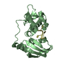
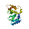
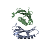
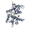
| Title | Crystal structure of the hyperthermophilic archaeal dna-binding protein Sso10b2 at 1.85 A | ||||||
 Components Components | DNA binding protein SSO10b | ||||||
 Keywords Keywords | DNA BINDING PROTEIN | ||||||
| Function / homology |  Function and homology information Function and homology informationchromosome condensation / chromosome / double-stranded DNA binding / RNA binding / cytoplasm Similarity search - Function | ||||||
| Biological species |   Sulfolobus solfataricus (archaea) Sulfolobus solfataricus (archaea) | ||||||
| Method |  X-RAY DIFFRACTION / SYNCHROTRON / MAD / Resolution: 1.85 Å X-RAY DIFFRACTION / SYNCHROTRON / MAD / Resolution: 1.85 Å | ||||||
 Authors Authors | Chou, C.-C. / Lin, T.-W. / Chen, C.-Y. / Wang, A.H.J. | ||||||
 Citation Citation | Journal: J.BACTERIOL. / Year: 2003 Title: Crystal structure of the hyperthermophilic archaeal DNA-binding protein Sso10b2 at a resolution of 1.85 Angstroms Authors: Chou, C.C. / Lin, T.W. / Chen, C.Y. / Wang, A.H.J. | ||||||
| History |
|
- Structure visualization
| Structure viewer | Molecule: MolmilJmol/JSmol |
|---|
- Downloads & links
-Download
| PDBx/mmCIF format | 1udv.cif.gz | 49.6 KB | Display | PDBx/mmCIF format |
|---|---|---|---|---|
| PDB format | pdb1udv.ent.gz | 36.4 KB | Display | PDB format |
| PDBx/mmJSON format | 1udv.json.gz | Tree view | PDBx/mmJSON format | |
| Others |  Other downloads Other downloads |
-Validation report
| Arichive directory | https://data.pdbj.org/pub/pdb/validation_reports/ud/1udvftp://data.pdbj.org/pub/pdb/validation_reports/ud/1udv | HTTPS FTP |
|---|
-Related structure data
| Similar structure data |
|---|


-Links
 PDBj
PDBj
- Assembly
| Deposited unit | 
| ||||||||
|---|---|---|---|---|---|---|---|---|---|
| 1 |
| ||||||||
| Unit cell |
| ||||||||
| Components on special symmetry positions |
|
-Components
| #1: Protein | Mass: 10255.882 Da / Num. of mol.: 2 Source method: isolated from a genetically manipulated source Source: (gene. exp.) Sulfolobus solfataricus (archaea) / Strain: P2 / Cellular location: CYTOPLASM / Gene: SSO6877 / Plasmid: PET30 / Species (production host): Escherichia coli / Cellular location (production host): CYTOPLASM / Production host:  #2: Chemical | ChemComp-ZN / |   Mass: 65.409 Da / Num. of mol.: 1 / Source method: obtained synthetically / Formula: Zn Mass: 65.409 Da / Num. of mol.: 1 / Source method: obtained synthetically / Formula: Zn#3: Water | ChemComp-HOH / |  Mass: 18.015 Da / Num. of mol.: 150 / Source method: isolated from a natural source / Formula: H2O Mass: 18.015 Da / Num. of mol.: 150 / Source method: isolated from a natural source / Formula: H2O |
|---|
-Experimental details
-Experiment
| Experiment | Method: X-RAY DIFFRACTION / Number of used crystals: 1 |
|---|
- Sample preparation
| Crystal | Density Matthews: 2.1 Å3/Da / Density % sol: 40 % | |||||||||||||||||||||||||||||||||||
|---|---|---|---|---|---|---|---|---|---|---|---|---|---|---|---|---|---|---|---|---|---|---|---|---|---|---|---|---|---|---|---|---|---|---|---|---|
| Crystal grow | Temperature: 298 K / Method: vapor diffusion / pH: 6.5 Details: PEG MME 550, ZnSO4, MES, pH 6.5, VAPOR DIFFUSION, temperature 298K | |||||||||||||||||||||||||||||||||||
| Crystal grow | *PLUS Method: vapor diffusion, hanging drop | |||||||||||||||||||||||||||||||||||
| Components of the solutions | *PLUS
|
-Data collection
| Diffraction | Mean temperature: 100 K |
|---|---|
| Diffraction source | Source: SYNCHROTRON / Site: NSRRC  / Beamline: BL17B2 / Wavelength: 1.1163 / Wavelength: 1.1163 Å / Beamline: BL17B2 / Wavelength: 1.1163 / Wavelength: 1.1163 Å |
| Detector | Type: RIGAKU RAXIS IV++ / Detector: IMAGE PLATE / Date: Jun 15, 2003 / Details: MIRROR |
| Radiation | Monochromator: NI FILTER / Protocol: SINGLE WAVELENGTH / Monochromatic (M) / Laue (L): M / Scattering type: x-ray |
| Radiation wavelength | Wavelength: 1.1163 Å / Relative weight: 1 |
| Reflection | Resolution: 1.85→31.78 Å / Num. all: 15639 / Num. obs: 15639 / % possible obs: 99.5 % / Observed criterion σ(F): 4 / Observed criterion σ(I): 2 / Redundancy: 4.5 % / Biso Wilson estimate: 19.6 Å2 / Rmerge(I) obs: 0.076 / Net I/σ(I): 15.9 |
| Reflection shell | Resolution: 1.85→1.92 Å / Redundancy: 3.8 % / Rmerge(I) obs: 0.229 / Mean I/σ(I) obs: 5.8 / Num. unique all: 1536 / % possible all: 97.6 |
| Reflection | *PLUS |
| Reflection shell | *PLUS % possible obs: 92.6 % |
- Processing
| Software |
| ||||||||||||||||||||||||||||||||||||||||||||||||||||||||||||||||||||||||||||||||
|---|---|---|---|---|---|---|---|---|---|---|---|---|---|---|---|---|---|---|---|---|---|---|---|---|---|---|---|---|---|---|---|---|---|---|---|---|---|---|---|---|---|---|---|---|---|---|---|---|---|---|---|---|---|---|---|---|---|---|---|---|---|---|---|---|---|---|---|---|---|---|---|---|---|---|---|---|---|---|---|---|---|
| Refinement | Method to determine structure: MAD / Resolution: 1.85→31.78 Å / Rfactor Rfree error: 0.01 / Data cutoff high rms absF: 1000000 / Isotropic thermal model: RESTRAINED / Cross valid method: THROUGHOUT / σ(F): 2
| ||||||||||||||||||||||||||||||||||||||||||||||||||||||||||||||||||||||||||||||||
| Solvent computation | Solvent model: FLAT MODEL / Bsol: 52.1562 Å2 / ksol: 0.354406 e/Å3 | ||||||||||||||||||||||||||||||||||||||||||||||||||||||||||||||||||||||||||||||||
| Displacement parameters | Biso mean: 34.8 Å2
| ||||||||||||||||||||||||||||||||||||||||||||||||||||||||||||||||||||||||||||||||
| Refine analyze |
| ||||||||||||||||||||||||||||||||||||||||||||||||||||||||||||||||||||||||||||||||
| Refinement step | Cycle: LAST / Resolution: 1.85→31.78 Å
| ||||||||||||||||||||||||||||||||||||||||||||||||||||||||||||||||||||||||||||||||
| Refine LS restraints |
| ||||||||||||||||||||||||||||||||||||||||||||||||||||||||||||||||||||||||||||||||
| LS refinement shell | Resolution: 1.85→1.97 Å / Rfactor Rfree error: 0.033 / Total num. of bins used: 6
| ||||||||||||||||||||||||||||||||||||||||||||||||||||||||||||||||||||||||||||||||
| Xplor file |
| ||||||||||||||||||||||||||||||||||||||||||||||||||||||||||||||||||||||||||||||||
| Refine LS restraints | *PLUS
|
