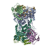
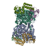
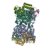
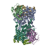
 Movie
Movie Controller


+ Open data

- Basic information
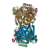
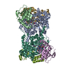
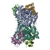
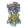
| Entry | Database: PDB / ID: 1uc4 | |||||||||
|---|---|---|---|---|---|---|---|---|---|---|
| Title | Structure of diol dehydratase complexed with (S)-1,2-propanediol | |||||||||
 Components Components | (diol dehydrase ...) x 3 | |||||||||
 Keywords Keywords | LYASE / ALPHA/BETA BARREL | |||||||||
| Function / homology |  Function and homology information Function and homology informationpropanediol dehydratase / propanediol dehydratase activity / cobalamin binding / metal ion binding Similarity search - Function | |||||||||
| Biological species |  Klebsiella oxytoca (bacteria) Klebsiella oxytoca (bacteria) | |||||||||
| Method |  X-RAY DIFFRACTION / SYNCHROTRON / MOLECULAR REPLACEMENT / Resolution: 1.8 Å X-RAY DIFFRACTION / SYNCHROTRON / MOLECULAR REPLACEMENT / Resolution: 1.8 Å | |||||||||
 Authors Authors | Shibata, N. / Nakanishi, Y. / Fukuoka, M. / Yamanishi, M. / Yasuoka, N. / Toraya, T. | |||||||||
 Citation Citation | Journal: J.BIOL.CHEM. / Year: 2003 Title: Structural rationalization for the lack of stereospecificity in coenzyme B12-dependent diol dehydratase Authors: Shibata, N. / Nakanishi, Y. / Fukuoka, M. / Yamanishi, M. / Yasuoka, N. / Toraya, T. | |||||||||
| History |
|
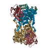
- Structure visualization
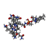
| Structure viewer | Molecule: MolmilJmol/JSmol |
|---|
- Downloads & links
-Download
| PDBx/mmCIF format | 1uc4.cif.gz | 383.7 KB | Display | PDBx/mmCIF format |
|---|---|---|---|---|
| PDB format | pdb1uc4.ent.gz | 303.2 KB | Display | PDB format |
| PDBx/mmJSON format | 1uc4.json.gz | Tree view | PDBx/mmJSON format | |
| Others |  Other downloads Other downloads |
-Validation report
| Arichive directory | https://data.pdbj.org/pub/pdb/validation_reports/uc/1uc4ftp://data.pdbj.org/pub/pdb/validation_reports/uc/1uc4 | HTTPS FTP |
|---|
-Related structure data
| Related structure data |  1uc5C 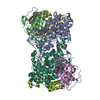 1egmS S: Starting model for refinement C: citing same article ( |
|---|---|
| Similar structure data |


-Links
 PDBj
PDBj
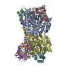
- Assembly
| Deposited unit | 
| ||||||||
|---|---|---|---|---|---|---|---|---|---|
| 1 |
| ||||||||
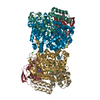
| Unit cell |
|
-Components
-Diol dehydrase ... , 3 types, 6 molecules ALBEGM
| #1: Protein | Mass: 60408.133 Da / Num. of mol.: 2 Source method: isolated from a genetically manipulated source Source: (gene. exp.) Klebsiella oxytoca (bacteria) / Plasmid: pUSI2E / Production host: #2: Protein | Mass: 24141.678 Da / Num. of mol.: 2 Source method: isolated from a genetically manipulated source Source: (gene. exp.) Klebsiella oxytoca (bacteria) / Plasmid: pUSI2E / Production host: #3: Protein | Mass: 19198.695 Da / Num. of mol.: 2 Source method: isolated from a genetically manipulated source Source: (gene. exp.) Klebsiella oxytoca (bacteria) / Plasmid: pUSI2E / Production host: |
|---|
-Non-polymers , 5 types, 1421 molecules 


| #4: Chemical | ChemComp-NH4 /  Mass: 18.038 Da / Num. of mol.: 4 / Source method: obtained synthetically / Formula: H4N Mass: 18.038 Da / Num. of mol.: 4 / Source method: obtained synthetically / Formula: H4N#5: Chemical |  Mass: 39.098 Da / Num. of mol.: 2 / Source method: obtained synthetically / Formula: K Mass: 39.098 Da / Num. of mol.: 2 / Source method: obtained synthetically / Formula: K#6: Chemical |  Mass: 76.094 Da / Num. of mol.: 2 / Source method: obtained synthetically / Formula: C3H8O2 Mass: 76.094 Da / Num. of mol.: 2 / Source method: obtained synthetically / Formula: C3H8O2#7: Chemical |  Mass: 1356.373 Da / Num. of mol.: 2 / Source method: obtained synthetically / Formula: C63H89CoN14O14P Mass: 1356.373 Da / Num. of mol.: 2 / Source method: obtained synthetically / Formula: C63H89CoN14O14P#8: Water | ChemComp-HOH / | Mass: 18.015 Da / Num. of mol.: 1411 / Source method: isolated from a natural source / Formula: H2O |
|---|
-Experimental details
-Experiment
| Experiment | Method: X-RAY DIFFRACTION / Number of used crystals: 1 |
|---|
- Sample preparation
| Crystal | Density Matthews: 2.25 Å3/Da / Density % sol: 44.82 % | ||||||||||||||||||||||||||||||||||||
|---|---|---|---|---|---|---|---|---|---|---|---|---|---|---|---|---|---|---|---|---|---|---|---|---|---|---|---|---|---|---|---|---|---|---|---|---|---|
| Crystal grow | Temperature: 278 K / Method: vapor diffusion / pH: 8 Details: PEG6000, ammonium sulfate, Tris, (S)-1,2,-propanediol, pH 8.0, VAPOR DIFFUSION, temperature 278K | ||||||||||||||||||||||||||||||||||||
| Crystal grow | *PLUS Temperature: 4 ℃ / Method: sandwich-drop vapor diffusion | ||||||||||||||||||||||||||||||||||||
| Components of the solutions | *PLUS
|
-Data collection
| Diffraction | Mean temperature: 100 K |
|---|---|
| Diffraction source | Source: SYNCHROTRON / Site: SPring-8  / Beamline: BL41XU / Wavelength: 0.708 Å / Beamline: BL41XU / Wavelength: 0.708 Å |
| Detector | Type: MARRESEARCH / Detector: CCD / Date: Apr 14, 2000 |
| Radiation | Protocol: SINGLE WAVELENGTH / Monochromatic (M) / Laue (L): M / Scattering type: x-ray |
| Radiation wavelength | Wavelength: 0.708 Å / Relative weight: 1 |
| Reflection | Resolution: 1.8→50 Å / % possible obs: 87.3 % / Observed criterion σ(F): 0 / Rmerge(I) obs: 0.055 |
| Reflection shell | Resolution: 1.8→1.86 Å / Rmerge(I) obs: 0.119 / % possible all: 0.671 |
| Reflection | *PLUS Highest resolution: 1.8 Å / Lowest resolution: 30 Å / Num. obs: 152278 / Num. measured all: 1991323 |
| Reflection shell | *PLUS % possible obs: 67.1 % |
- Processing
| Software |
| |||||||||||||||||||||||||||||||||||||||||||||||||
|---|---|---|---|---|---|---|---|---|---|---|---|---|---|---|---|---|---|---|---|---|---|---|---|---|---|---|---|---|---|---|---|---|---|---|---|---|---|---|---|---|---|---|---|---|---|---|---|---|---|---|
| Refinement | Method to determine structure: MOLECULAR REPLACEMENT Starting model: PDB ENTRY 1EGM Resolution: 1.8→36.92 Å / Cross valid method: THROUGHOUT / σ(F): 0 / Stereochemistry target values: Engh & Huber
| |||||||||||||||||||||||||||||||||||||||||||||||||
| Refine analyze |
| |||||||||||||||||||||||||||||||||||||||||||||||||
| Refinement step | Cycle: LAST / Resolution: 1.8→36.92 Å
| |||||||||||||||||||||||||||||||||||||||||||||||||
| Refine LS restraints |
| |||||||||||||||||||||||||||||||||||||||||||||||||
| LS refinement shell |
| |||||||||||||||||||||||||||||||||||||||||||||||||
| Refinement | *PLUS Lowest resolution: 30 Å / Rfactor Rfree: 0.23 | |||||||||||||||||||||||||||||||||||||||||||||||||
| Solvent computation | *PLUS | |||||||||||||||||||||||||||||||||||||||||||||||||
| Displacement parameters | *PLUS | |||||||||||||||||||||||||||||||||||||||||||||||||
| Refine LS restraints | *PLUS
|
