 Movie
Movie Controller

[English] 日本語
 Yorodumi
- PDB-1egm: CRYSTAL STRUCTURE OF DIOL DEHYDRATASE-CYANOCOBALAMIN COMPLEX AT 100K. -
+ Open data

- Basic information
| Entry | Database: PDB / ID: 1egm | |||||||||
|---|---|---|---|---|---|---|---|---|---|---|
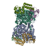
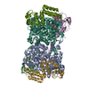
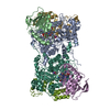
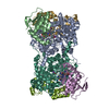
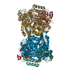
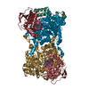
| Title | CRYSTAL STRUCTURE OF DIOL DEHYDRATASE-CYANOCOBALAMIN COMPLEX AT 100K. | |||||||||
 Components Components | (PROPANEDIOL DEHYDRATASE) x 3 | |||||||||
 Keywords Keywords | LYASE / CYANOCOBALAMIN / PROPANEDIOL / POTASSIUM ION / TIM BARREL | |||||||||
| Function / homology |  Function and homology information Function and homology informationpropanediol dehydratase / propanediol dehydratase activity / cobalamin binding / metal ion binding Similarity search - Function | |||||||||
| Biological species |  Klebsiella oxytoca (bacteria) Klebsiella oxytoca (bacteria) | |||||||||
| Method |  X-RAY DIFFRACTION / SYNCHROTRON / Resolution: 1.85 Å X-RAY DIFFRACTION / SYNCHROTRON / Resolution: 1.85 Å | |||||||||
 Authors Authors | Masuda, J. / Shibata, N. / Toraya, T. / Morimoto, Y. / Yasuoka, N. | |||||||||
 Citation Citation | Journal: Structure Fold.Des. / Year: 2000 Title: How a protein generates a catalytic radical from coenzyme B(12): X-ray structure of a diol-dehydratase-adeninylpentylcobalamin complex. Authors: Masuda, J. / Shibata, N. / Morimoto, Y. / Toraya, T. / Yasuoka, N. #1: Journal: Structure / Year: 1999Title: A New Mode of B12 Binding and the Direct Participation of a Potassium Ion in Enzyme Catalysis: X-ray Structure of Diol Dehydratase Authors: Shibata, N. / Masuda, J. / Toraya, T. / Morimoto, Y. / Yasuoka, N. | |||||||||
| History |
|
- Structure visualization
| Structure viewer | Molecule: MolmilJmol/JSmol |
|---|
- Downloads & links
-Download
| PDBx/mmCIF format | 1egm.cif.gz | 378.8 KB | Display | PDBx/mmCIF format |
|---|---|---|---|---|
| PDB format | pdb1egm.ent.gz | 301.8 KB | Display | PDB format |
| PDBx/mmJSON format | 1egm.json.gz | Tree view | PDBx/mmJSON format | |
| Others |  Other downloads Other downloads |
-Validation report
| Arichive directory | https://data.pdbj.org/pub/pdb/validation_reports/eg/1egmftp://data.pdbj.org/pub/pdb/validation_reports/eg/1egm | HTTPS FTP |
|---|
-Related structure data


-Links
 PDBj
PDBj- Assembly
| Deposited unit | 
| ||||||||
|---|---|---|---|---|---|---|---|---|---|
| 1 |
| ||||||||
| Unit cell |
| ||||||||
| Details | The biological assembly is heterohexamer constructed from A, B, G, L, E, G chains and two cyanocobalamins. |
-Components
-Protein , 3 types, 6 molecules ALBEGM
| #1: Protein | Mass: 60408.133 Da / Num. of mol.: 2 / Fragment: ALPHA CHAIN Source method: isolated from a genetically manipulated source Source: (gene. exp.) Klebsiella oxytoca (bacteria) / Plasmid: PUC119 / Production host: #2: Protein | Mass: 24141.678 Da / Num. of mol.: 2 / Fragment: BETA CHAIN Source method: isolated from a genetically manipulated source Source: (gene. exp.) Klebsiella oxytoca (bacteria) / Plasmid: PUC119 / Production host: #3: Protein | Mass: 19198.695 Da / Num. of mol.: 2 / Fragment: GAMMA CHAIN Source method: isolated from a genetically manipulated source Source: (gene. exp.) Klebsiella oxytoca (bacteria) / Plasmid: PUC119 / Production host: |
|---|
-Non-polymers , 4 types, 1217 molecules 


| #4: Chemical |  Mass: 39.098 Da / Num. of mol.: 2 / Source method: obtained synthetically / Formula: K Mass: 39.098 Da / Num. of mol.: 2 / Source method: obtained synthetically / Formula: K#5: Chemical |  Mass: 1356.373 Da / Num. of mol.: 2 / Source method: obtained synthetically / Formula: C63H89CoN14O14P Mass: 1356.373 Da / Num. of mol.: 2 / Source method: obtained synthetically / Formula: C63H89CoN14O14P#6: Chemical |  Mass: 76.094 Da / Num. of mol.: 2 / Source method: obtained synthetically / Formula: C3H8O2 Mass: 76.094 Da / Num. of mol.: 2 / Source method: obtained synthetically / Formula: C3H8O2#7: Water | ChemComp-HOH / | Mass: 18.015 Da / Num. of mol.: 1211 / Source method: isolated from a natural source / Formula: H2O |
|---|
-Experimental details
-Experiment
| Experiment | Method: X-RAY DIFFRACTION / Number of used crystals: 1 |
|---|
- Sample preparation
| Crystal | Density Matthews: 2.25 Å3/Da / Density % sol: 45.35 % | |||||||||||||||||||||||||
|---|---|---|---|---|---|---|---|---|---|---|---|---|---|---|---|---|---|---|---|---|---|---|---|---|---|---|
| Crystal grow | Temperature: 277 K / Method: vapor diffusion, sandwich / pH: 8 Details: PEG6000, potassium phosphate, Tris, lauryldimethylamine oxide, ammonium sulfate, propane diol, cyanocobalamin, pH 8.0, VAPOR DIFFUSION, SANDWICH, temperature 277K | |||||||||||||||||||||||||
| Crystal grow | *PLUS Details: Masuda, J., (1999) Acta Crystallogr, D55, 907. / Method: vapor diffusion, sandwich drop | |||||||||||||||||||||||||
| Components of the solutions | *PLUS
|
-Data collection
| Diffraction | Mean temperature: 100 K |
|---|---|
| Diffraction source | Source: SYNCHROTRON / Site: SPring-8  / Beamline: BL41XU / Wavelength: 1 / Beamline: BL41XU / Wavelength: 1 |
| Detector | Type: MARRESEARCH / Detector: CCD / Date: Oct 3, 1999 |
| Radiation | Protocol: SINGLE WAVELENGTH / Monochromatic (M) / Laue (L): M / Scattering type: x-ray |
| Radiation wavelength | Wavelength: 1 Å / Relative weight: 1 |
| Reflection | Resolution: 1.85→40 Å / Num. all: 142052 / Num. obs: 142006 / % possible obs: 88.6 % / Observed criterion σ(F): 1 / Observed criterion σ(I): 1 / Redundancy: 5.4 % / Biso Wilson estimate: 19.8 Å2 / Rmerge(I) obs: 0.072 / Net I/σ(I): 21.9 |
| Reflection shell | Resolution: 1.85→1.88 Å / Redundancy: 2.6 % / Rmerge(I) obs: 0.134 / Num. unique all: 5330 / % possible all: 67.7 |
| Reflection | *PLUS Num. obs: 142053 / Num. measured all: 1939544 |
| Reflection shell | *PLUS % possible obs: 67.7 % |
- Processing
| Software |
| ||||||||||||||||||||
|---|---|---|---|---|---|---|---|---|---|---|---|---|---|---|---|---|---|---|---|---|---|
| Refinement | Resolution: 1.85→30 Å / σ(F): 1 / σ(I): 1 / Stereochemistry target values: Engh & Huber
| ||||||||||||||||||||
| Refinement step | Cycle: LAST / Resolution: 1.85→30 Å
| ||||||||||||||||||||
| Refine LS restraints |
| ||||||||||||||||||||
| Software | *PLUS Name: SHELXL-97 / Classification: refinement | ||||||||||||||||||||
| Refinement | *PLUS Rfactor Rfree: 0.243 / Rfactor Rwork: 0.182 | ||||||||||||||||||||
| Solvent computation | *PLUS | ||||||||||||||||||||
| Displacement parameters | *PLUS |
