 Movie
Movie Controller


+ Open data

- Basic information
| Entry | Database: PDB / ID: 1mmf | ||||||
|---|---|---|---|---|---|---|---|
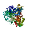
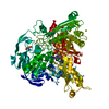
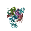
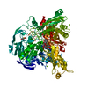
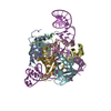
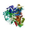
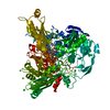
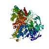
| Title | Crystal structure of substrate free form of glycerol dehydratase | ||||||
 Components Components | (glycerol dehydrase ...) x 3 | ||||||
 Keywords Keywords | LYASE / Glycerol dehydratase / Diol dehydratase / Coenzyme B12 / TIM barrel | ||||||
| Function / homology |  Function and homology information Function and homology informationglycerol dehydratase / glycerol dehydratase activity / propanediol dehydratase / propanediol dehydratase activity / cobalamin binding Similarity search - Function | ||||||
| Biological species |  Klebsiella pneumoniae (bacteria) Klebsiella pneumoniae (bacteria) | ||||||
| Method |  X-RAY DIFFRACTION / SYNCHROTRON / MOLECULAR REPLACEMENT / Resolution: 2.5 Å X-RAY DIFFRACTION / SYNCHROTRON / MOLECULAR REPLACEMENT / Resolution: 2.5 Å | ||||||
 Authors Authors | Liao, D.I. / Dotson, G. / Turner, I. / Reiss, L. / Emptage, M. | ||||||
 Citation Citation | Journal: J.Inorg.Biochem. / Year: 2003 Title: Crystal structure of substrate free form of glycerol dehydratase Authors: Liao, D.I. / Dotson, G. / Turner, I. / Reiss, L. / Emptage, M. | ||||||
| History |
|
- Structure visualization
| Structure viewer | Molecule: MolmilJmol/JSmol |
|---|
- Downloads & links
-Download
| PDBx/mmCIF format | 1mmf.cif.gz | 345.9 KB | Display | PDBx/mmCIF format |
|---|---|---|---|---|
| PDB format | pdb1mmf.ent.gz | 277.5 KB | Display | PDB format |
| PDBx/mmJSON format | 1mmf.json.gz | Tree view | PDBx/mmJSON format | |
| Others |  Other downloads Other downloads |
-Validation report
| Arichive directory | https://data.pdbj.org/pub/pdb/validation_reports/mm/1mmfftp://data.pdbj.org/pub/pdb/validation_reports/mm/1mmf | HTTPS FTP |
|---|
-Related structure data
| Related structure data | 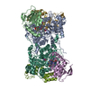 1dioS S: Starting model for refinement |
|---|---|
| Similar structure data |

-Links
 PDBj
PDBj- Assembly
| Deposited unit | 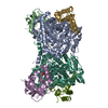
| ||||||||
|---|---|---|---|---|---|---|---|---|---|
| 1 | 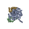
| ||||||||
| 2 | 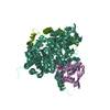
| ||||||||
| 3 |
| ||||||||
| Unit cell |
| ||||||||
| Details | The biologyical assembly of glycerol dehydratase is a heterohexamer. It is a dimer of alpha-beta-gamma trimers. The asymmetric unit in the crystal contains an entire heterohexamer. Chain names for subunits are A, B, and G fare one trimer and L, E and M for the second trimer. |
-Components
-Glycerol dehydrase ... , 3 types, 6 molecules ALBEGM
| #1: Protein | Mass: 60714.555 Da / Num. of mol.: 2 Source method: isolated from a genetically manipulated source Source: (gene. exp.) Klebsiella pneumoniae (bacteria) / Plasmid: pBluescript SKII / Species (production host): Escherichia coli / Production host: #2: Protein | Mass: 21353.424 Da / Num. of mol.: 2 Source method: isolated from a genetically manipulated source Source: (gene. exp.) Klebsiella pneumoniae (bacteria) / Plasmid: pBluescript SKII / Species (production host): Escherichia coli / Production host: #3: Protein | Mass: 16128.362 Da / Num. of mol.: 2 Source method: isolated from a genetically manipulated source Source: (gene. exp.) Klebsiella pneumoniae (bacteria) / Plasmid: pBluescript SKII / Species (production host): Escherichia coli / Production host: |
|---|
-Non-polymers , 3 types, 211 molecules 


| #4: Chemical |  Mass: 39.098 Da / Num. of mol.: 2 / Source method: obtained synthetically / Formula: K Mass: 39.098 Da / Num. of mol.: 2 / Source method: obtained synthetically / Formula: K#5: Chemical |  Mass: 1330.356 Da / Num. of mol.: 2 / Source method: obtained synthetically / Formula: C62H89CoN13O14P Mass: 1330.356 Da / Num. of mol.: 2 / Source method: obtained synthetically / Formula: C62H89CoN13O14P#6: Water | ChemComp-HOH / | Mass: 18.015 Da / Num. of mol.: 207 / Source method: isolated from a natural source / Formula: H2O |
|---|
-Experimental details
-Experiment
| Experiment | Method: X-RAY DIFFRACTION / Number of used crystals: 1 |
|---|
- Sample preparation
| Crystal | Density Matthews: 2.87 Å3/Da / Density % sol: 57.12 % | ||||||||||||||||||||||||||||||||||||||||||||||||||||||||
|---|---|---|---|---|---|---|---|---|---|---|---|---|---|---|---|---|---|---|---|---|---|---|---|---|---|---|---|---|---|---|---|---|---|---|---|---|---|---|---|---|---|---|---|---|---|---|---|---|---|---|---|---|---|---|---|---|---|
| Crystal grow | Temperature: 298 K / Method: vapor diffusion, hanging drop / pH: 9.4 Details: Amonium sulfate, RbCl2, CdCl2, cyanocobalamin, CHES, pH 9.4, VAPOR DIFFUSION, HANGING DROP, temperature 298K | ||||||||||||||||||||||||||||||||||||||||||||||||||||||||
| Crystal grow | *PLUS pH: 7.5 / Method: vapor diffusion, hanging drop | ||||||||||||||||||||||||||||||||||||||||||||||||||||||||
| Components of the solutions | *PLUS
|
-Data collection
| Diffraction | Mean temperature: 100 K |
|---|---|
| Diffraction source | Source: SYNCHROTRON / Site: APS  / Beamline: 5ID-B / Wavelength: 1 Å / Beamline: 5ID-B / Wavelength: 1 Å |
| Detector | Type: MARRESEARCH / Detector: CCD / Date: Aug 11, 1999 |
| Radiation | Protocol: SINGLE WAVELENGTH / Monochromatic (M) / Laue (L): M / Scattering type: x-ray |
| Radiation wavelength | Wavelength: 1 Å / Relative weight: 1 |
| Reflection | Resolution: 2.5→30 Å / Num. all: 67426 / Num. obs: 67426 / % possible obs: 87.7 % / Observed criterion σ(I): -3 / Rmerge(I) obs: 0.072 / Net I/σ(I): 11.6 |
| Reflection shell | Resolution: 2.5→2.54 Å / Rmerge(I) obs: 0.14 / Mean I/σ(I) obs: 3.1 / % possible all: 50.1 |
| Reflection | *PLUS Lowest resolution: 30 Å / Num. measured all: 302300 |
| Reflection shell | *PLUS Highest resolution: 2.5 Å / % possible obs: 50.1 % / Rmerge(I) obs: 0.14 |
- Processing
| Software |
| |||||||||||||||
|---|---|---|---|---|---|---|---|---|---|---|---|---|---|---|---|---|
| Refinement | Method to determine structure: MOLECULAR REPLACEMENT Starting model: PDB ENTRY 1DIO Resolution: 2.5→30 Å / σ(F): 2 / Stereochemistry target values: Engh & Huber
| |||||||||||||||
| Refinement step | Cycle: LAST / Resolution: 2.5→30 Å
| |||||||||||||||
| Refine LS restraints |
| |||||||||||||||
| Refinement | *PLUS % reflection Rfree: 5 % | |||||||||||||||
| Solvent computation | *PLUS | |||||||||||||||
| Displacement parameters | *PLUS |
