 Movie
Movie Controller

[English] 日本語
 Yorodumi
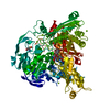
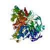
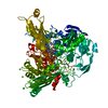
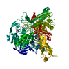
- PDB-1vlb: STRUCTURE REFINEMENT OF THE ALDEHYDE OXIDOREDUCTASE FROM DESULFOV... -
+ Open data

- Basic information
| Entry | Database: PDB / ID: 1vlb | |||||||||
|---|---|---|---|---|---|---|---|---|---|---|
| Title | STRUCTURE REFINEMENT OF THE ALDEHYDE OXIDOREDUCTASE FROM DESULFOVIBRIO GIGAS AT 1.28 A | |||||||||
 Components Components | ALDEHYDE OXIDOREDUCTASE | |||||||||
 Keywords Keywords | OXIDOREDUCTASE / Aldehyde Oxidoreductase / Desulfovibrio Gigas / iron-sulphur cluster | |||||||||
| Function / homology |  Function and homology information Function and homology informationaldehyde dehydrogenase (FAD-independent) / aldehyde dehydrogenase (FAD-independent) activity / 2 iron, 2 sulfur cluster binding / iron ion binding Similarity search - Function | |||||||||
| Biological species |  Desulfovibrio gigas (bacteria) Desulfovibrio gigas (bacteria) | |||||||||
| Method |  X-RAY DIFFRACTION / SYNCHROTRON / MOLECULAR REPLACEMENT / Resolution: 1.28 Å X-RAY DIFFRACTION / SYNCHROTRON / MOLECULAR REPLACEMENT / Resolution: 1.28 Å | |||||||||
 Authors Authors | Rebelo, J.M. / Dias, J.M. / Huber, R. / Moura, J.J.G. / Romao, M.J. | |||||||||
 Citation Citation | Journal: J.Biol.Inorg.Chem. / Year: 2001 Title: Structure refinement of the aldehyde oxidoreductase from Desulfovibrio gigas (MOP) at 1.28 A Authors: Rebelo, J.M. / Dias, J.M. / Huber, R. / Moura, J.J.G. / Romao, M.J. #1: Journal: Science / Year: 1995Title: Structure of the Aldehyde Oxido-reductase from Desulfovibrio gigas at 2.25 A Resolution: A Member of the Xanthine Oxidase Protein Family Authors: Romao, M.J. / Archer, M. / Moura, I. / Moura, J.J.G. / LeGall, J. / Engh, R. / Schneider, M. / Hof, P. / Huber, R. #2: Journal: Proc.Natl.Acad.Sci.USA / Year: 1996Title: A structure-based catalytic mechanism for the xanthine oxidase family of molybdenum enzymes Authors: Huber, R. / Hof, P. / Duarte, R.O. / Moura, J.J.G. / Moura, I. / Liu, M.-Y. / LeGall, J. / Hille, R. / Archer, M. / Romao, M.J. | |||||||||
| History |
|
- Structure visualization
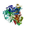
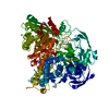
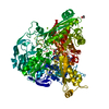
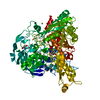
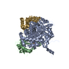
| Structure viewer | Molecule: MolmilJmol/JSmol |
|---|
- Downloads & links
-Download
| PDBx/mmCIF format | 1vlb.cif.gz | 414.8 KB | Display | PDBx/mmCIF format |
|---|---|---|---|---|
| PDB format | pdb1vlb.ent.gz | 335.6 KB | Display | PDB format |
| PDBx/mmJSON format | 1vlb.json.gz | Tree view | PDBx/mmJSON format | |
| Others |  Other downloads Other downloads |
-Validation report
| Arichive directory | https://data.pdbj.org/pub/pdb/validation_reports/vl/1vlbftp://data.pdbj.org/pub/pdb/validation_reports/vl/1vlb | HTTPS FTP |
|---|
-Related structure data
| Related structure data |  1alo S: Starting model for refinement |
|---|---|
| Similar structure data |


-Links
 PDBj
PDBj


- Assembly
| Deposited unit | 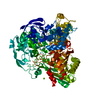
| ||||||||||
|---|---|---|---|---|---|---|---|---|---|---|---|
| 1 |
| ||||||||||
| Unit cell |
| ||||||||||
| Components on special symmetry positions |
|
-Components
-Protein , 1 types, 1 molecules A
| #1: Protein | Mass: 97140.508 Da / Num. of mol.: 1 / Source method: isolated from a natural source / Source: (natural) Desulfovibrio gigas (bacteria) / References: UniProt: Q46509, aldehyde oxidase |
|---|
-Non-polymers , 6 types, 1248 molecules 


| #2: Chemical |  Mass: 35.453 Da / Num. of mol.: 3 / Source method: obtained synthetically / Formula: Cl Mass: 35.453 Da / Num. of mol.: 3 / Source method: obtained synthetically / Formula: Cl#3: Chemical |  Mass: 24.305 Da / Num. of mol.: 2 / Source method: obtained synthetically / Formula: Mg Mass: 24.305 Da / Num. of mol.: 2 / Source method: obtained synthetically / Formula: Mg#4: Chemical | ChemComp-PCD / ( |  Mass: 844.471 Da / Num. of mol.: 1 / Source method: obtained synthetically / Formula: C19H26MoN8O16P2S2 Mass: 844.471 Da / Num. of mol.: 1 / Source method: obtained synthetically / Formula: C19H26MoN8O16P2S2#5: Chemical |  Mass: 175.820 Da / Num. of mol.: 2 / Source method: obtained synthetically / Formula: Fe2S2 Mass: 175.820 Da / Num. of mol.: 2 / Source method: obtained synthetically / Formula: Fe2S2#6: Chemical |  Mass: 60.095 Da / Num. of mol.: 2 / Source method: obtained synthetically / Formula: C3H8O Mass: 60.095 Da / Num. of mol.: 2 / Source method: obtained synthetically / Formula: C3H8O#7: Water | ChemComp-HOH / | Mass: 18.015 Da / Num. of mol.: 1238 / Source method: isolated from a natural source / Formula: H2O |
|---|
-Experimental details
-Experiment
| Experiment | Method: X-RAY DIFFRACTION / Number of used crystals: 1 |
|---|
- Sample preparation
| Crystal | Density Matthews: 2.4 Å3/Da / Density % sol: 48.77 % |
|---|---|
| Crystal grow | Temperature: 277 K / Method: vapor diffusion, sitting drop / pH: 7.5 Details: MgCl2, isopropyl alcohol, pH 7.5, VAPOR DIFFUSION, SITTING DROP at 277K |
-Data collection
| Diffraction | Mean temperature: 100 K |
|---|---|
| Diffraction source | Source: SYNCHROTRON / Site: MPG/DESY, HAMBURG  / Beamline: BW6 / Wavelength: 1.0004 Å / Beamline: BW6 / Wavelength: 1.0004 Å |
| Detector | Type: MARRESEARCH / Detector: IMAGE PLATE / Date: Nov 7, 1998 |
| Radiation | Monochromator: double crystal monochromator / Protocol: SINGLE WAVELENGTH / Monochromatic (M) / Laue (L): M / Scattering type: x-ray |
| Radiation wavelength | Wavelength: 1.0004 Å / Relative weight: 1 |
| Reflection | Resolution: 1.28→24.4 Å / Num. all: 233755 / Num. obs: 226751 / % possible obs: 93 % / Observed criterion σ(I): 2 / Redundancy: 2 % / Biso Wilson estimate: 16.2 Å2 / Rmerge(I) obs: 0.091 / Rsym value: 0.036 / Net I/σ(I): 17.7 |
| Reflection shell | Resolution: 1.28→1.33 Å / Redundancy: 2 % / Rmerge(I) obs: 0.649 / Mean I/σ(I) obs: 1.6 / Rsym value: 0.436 / % possible all: 92.3 |
- Processing
| Software |
| |||||||||||||||||||||||||
|---|---|---|---|---|---|---|---|---|---|---|---|---|---|---|---|---|---|---|---|---|---|---|---|---|---|---|
| Refinement | Method to determine structure: MOLECULAR REPLACEMENT Starting model: PDB ENTRY 1ALO 1alo Resolution: 1.28→24.4 Å / Isotropic thermal model: anisotropic / Cross valid method: THROUGHOUT / σ(F): 4 / σ(I): 2 / Stereochemistry target values: Engh & Huber / Details: Used weighted full matrix least squares procedure
| |||||||||||||||||||||||||
| Displacement parameters | Biso mean: 24.7 Å2 | |||||||||||||||||||||||||
| Refine analyze | Luzzati coordinate error obs: 0.1 Å | |||||||||||||||||||||||||
| Refinement step | Cycle: LAST / Resolution: 1.28→24.4 Å
| |||||||||||||||||||||||||
| Refine LS restraints |
|
