 Movie
Movie Controller

[English] 日本語
 Yorodumi
- PDB-1trj: Homology Model of Yeast RACK1 Protein fitted into 11.7A cryo-EM m... -
+ Open data

- Basic information
| Entry | Database: PDB / ID: 1trj | ||||||
|---|---|---|---|---|---|---|---|
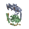
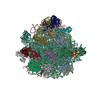
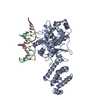
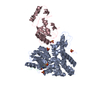
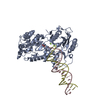
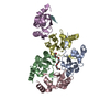
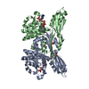
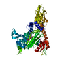
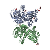
| Title | Homology Model of Yeast RACK1 Protein fitted into 11.7A cryo-EM map of Yeast 80S Ribosome | ||||||
 Components Components |
| ||||||
 Keywords Keywords | SIGNALING PROTEIN / RACK1 / Ribosome / homology model | ||||||
| Function / homology |  Function and homology information Function and homology informationregulation of amino acid metabolic process / negative regulation of glucose mediated signaling pathway / ribosome-associated ubiquitin-dependent protein catabolic process / GDP-dissociation inhibitor activity / nonfunctional rRNA decay / mRNA destabilization / negative regulation of translational frameshifting / G-protein alpha-subunit binding / translation regulator activity / rescue of stalled cytosolic ribosome ...regulation of amino acid metabolic process / negative regulation of glucose mediated signaling pathway / ribosome-associated ubiquitin-dependent protein catabolic process / GDP-dissociation inhibitor activity / nonfunctional rRNA decay / mRNA destabilization / negative regulation of translational frameshifting / G-protein alpha-subunit binding / translation regulator activity / rescue of stalled cytosolic ribosome / protein kinase C binding / maintenance of translational fidelity / ribosome binding / cytosolic small ribosomal subunit / cytoplasmic translation / negative regulation of translation / G protein-coupled receptor signaling pathway / negative regulation of gene expression / nucleus / cytoplasm / cytosol Similarity search - Function | ||||||
| Biological species |  | ||||||
| Method | ELECTRON MICROSCOPY / single particle reconstruction / cryo EM / Resolution: 11.7 Å | ||||||
 Authors Authors | Sengupta, J. / Nilsson, J. / Gursky, R. / Spahn, C.M. / Nissen, P. / Frank, J. | ||||||
 Citation Citation | Journal: Nat Struct Mol Biol / Year: 2004 Title: Identification of the versatile scaffold protein RACK1 on the eukaryotic ribosome by cryo-EM. Authors: Jayati Sengupta / Jakob Nilsson / Richard Gursky / Christian M T Spahn / Poul Nissen / Joachim Frank /  Abstract: RACK1 serves as a scaffold protein for a wide range of kinases and membrane-bound receptors. It is a WD-repeat family protein and is predicted to have a beta-propeller architecture with seven blades ...RACK1 serves as a scaffold protein for a wide range of kinases and membrane-bound receptors. It is a WD-repeat family protein and is predicted to have a beta-propeller architecture with seven blades like a Gbeta protein. Mass spectrometry studies have identified its association with the small subunit of eukaryotic ribosomes and, most recently, it has been shown to regulate initiation by recruiting protein kinase C to the 40S subunit. Here we present the results of a cryo-EM study of the 80S ribosome that positively locate RACK1 on the head region of the 40S subunit, in the immediate vicinity of the mRNA exit channel. One face of RACK1 exposes the WD-repeats as a platform for interactions with kinases and receptors. Using this platform, RACK1 can recruit other proteins to the ribosome. #1: Journal: Nature / Year: 2003Title: Release of eIF6 (p27BBP) from the 60S subunit allows 80S ribosome assembly Authors: Ceci, M. / Gaviraghi, C. / Gorrini, C. / Sala, L.A. / Offenhauser, N. / Marchisio, P.C. / Biffo, S. #2: Journal: J.Biol.Chem. / Year: 2003Title: Cpc2/RACK1 is a ribosome-associated protein that promotes efficient translation in Schizosaccharomyces pombe Authors: Shor, B. / Calaycay, J. / Rushbrook, J. / McLeod, M. #3: Journal: Biochem.J. / Year: 2004Title: Asc1p, a WD40-domain containing adaptor protein, is required for the interaction of the RNA-binding protein Scp160p with polysomes Authors: Baum, S. / Bittins, M. / Frey, S. / Seedorf, M. | ||||||
| History |
| ||||||
| Remark 999 | SEQUENCE ONLY COORDINATES FOR CA ATOMS FROM THE PROTEIN AND P ATOMS FROM THE NUCLEIC ACIDS WERE ...SEQUENCE ONLY COORDINATES FOR CA ATOMS FROM THE PROTEIN AND P ATOMS FROM THE NUCLEIC ACIDS WERE INCLUDED IN THE STRUCTURE. |
- Structure visualization
| Movie |
Movie viewer |
|---|---|
| Structure viewer | Molecule: MolmilJmol/JSmol |
- Downloads & links
-Download
| PDBx/mmCIF format | 1trj.cif.gz | 26.5 KB | Display | PDBx/mmCIF format |
|---|---|---|---|---|
| PDB format | pdb1trj.ent.gz | 10.5 KB | Display | PDB format |
| PDBx/mmJSON format | 1trj.json.gz | Tree view | PDBx/mmJSON format | |
| Others |  Other downloads Other downloads |
-Validation report
| Arichive directory | https://data.pdbj.org/pub/pdb/validation_reports/tr/1trjftp://data.pdbj.org/pub/pdb/validation_reports/tr/1trj | HTTPS FTP |
|---|
-Related structure data
| Related structure data | 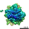 1067M M: map data used to model this data |
|---|---|
| Similar structure data |

-Links
 PDBj
PDBj


- Assembly
| Deposited unit | 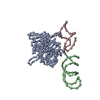
|
|---|---|
| 1 |
|
-Components
| #1: RNA chain | Mass: 13155.822 Da / Num. of mol.: 1 / Source method: obtained synthetically |
|---|---|
| #2: RNA chain | Mass: 8868.415 Da / Num. of mol.: 1 / Source method: obtained synthetically |
| #3: Protein | Mass: 34324.609 Da / Num. of mol.: 1 / Source method: isolated from a natural source / Source: (natural) |
-Experimental details
-Experiment
| Experiment | Method: ELECTRON MICROSCOPY |
|---|---|
| EM experiment | Aggregation state: PARTICLE / 3D reconstruction method: single particle reconstruction |
- Sample preparation
| Component | Name: 80S ribosome / Type: RIBOSOME |
|---|---|
| Buffer solution | pH: 7.5 |
| Specimen | Conc.: 32 mg/ml / Embedding applied: NO / Shadowing applied: NO / Staining applied: NO / Vitrification applied: YES |
| Specimen support | Details: QUANTIFOIL HOLLEY CARBON FILM GRIDS |
| Vitrification | Cryogen name: ETHANE / Details: RAPID-FREEZING IN LIQUID ETHANE |
- Electron microscopy imaging
| Microscopy | Model: FEI TECNAI 20 / Date: Feb 1, 2001 |
|---|---|
| Electron gun | Electron source:  FIELD EMISSION GUN / Accelerating voltage: 200 kV / Illumination mode: FLOOD BEAM FIELD EMISSION GUN / Accelerating voltage: 200 kV / Illumination mode: FLOOD BEAM |
| Electron lens | Mode: BRIGHT FIELD / Nominal magnification: 50000 X / Calibrated magnification: 52000 X / Nominal defocus max: 1400 nm / Nominal defocus min: 4900 nm |
| Specimen holder | Temperature: 93 K / Tilt angle max: 0 ° / Tilt angle min: 0 ° |
| Image recording | Electron dose: 15 e/Å2 / Film or detector model: KODAK SO-163 FILM |
- Processing
| EM software | Name: SPIDER / Category: 3D reconstruction | ||||||||||||
|---|---|---|---|---|---|---|---|---|---|---|---|---|---|
| CTF correction | Details: CTF CORRECTION OF 3D MAPS BY WIENER FILTERATION | ||||||||||||
| Symmetry | Point symmetry: C1 (asymmetric) | ||||||||||||
| 3D reconstruction | Method: 3D PROJECTION MATCHING GRADIENT WITH REGULARIZATION / Resolution: 11.7 Å / Actual pixel size: 2.93 Å / Magnification calibration: TMV / Details: SPIDER PACKAGE / Symmetry type: POINT | ||||||||||||
| Atomic model building | Protocol: OTHER / Space: REAL Details: METHOD--USING A RAPID MOTIF SEARCH MODULE OF SPIDER (THE CRYO-EM MAP USED FOR FITTING WAS INTERPOLATED TO PIXEL SIZE 2.82) | ||||||||||||
| Refinement | Highest resolution: 11.7 Å | ||||||||||||
| Refinement step | Cycle: LAST / Highest resolution: 11.7 Å
|
