 Movie
Movie Controller

[English] 日本語
 Yorodumi
- PDB-4v4b: Structure of the ribosomal 80S-eEF2-sordarin complex from yeast o... -
+ Open data

- Basic information
| Entry | Database: PDB / ID: 4v4b | |||||||||
|---|---|---|---|---|---|---|---|---|---|---|
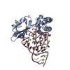
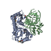
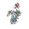
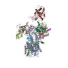
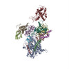
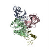
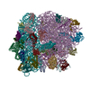
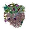
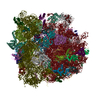
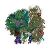
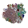
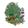
| Title | Structure of the ribosomal 80S-eEF2-sordarin complex from yeast obtained by docking atomic models for RNA and protein components into a 11.7 A cryo-EM map. | |||||||||
 Components Components |
| |||||||||
 Keywords Keywords | RIBOSOME / 80S ribosome / 40S ribosomal subunit / eEF2 / tRNA translocation / sordarin / cryo-EM | |||||||||
| Function / homology |  Function and homology information Function and homology information: / Ribosome Quality Control (RQC) complex extracts and degrades nascent peptide / PELO:HBS1L and ABCE1 dissociate a ribosome on a non-stop mRNA / Peptide chain elongation / Synthesis of diphthamide-EEF2 / positive regulation of translational elongation / ribophagy / positive regulation of translational fidelity / RMTs methylate histone arginines / Protein methylation ...: / Ribosome Quality Control (RQC) complex extracts and degrades nascent peptide / PELO:HBS1L and ABCE1 dissociate a ribosome on a non-stop mRNA / Peptide chain elongation / Synthesis of diphthamide-EEF2 / positive regulation of translational elongation / ribophagy / positive regulation of translational fidelity / RMTs methylate histone arginines / Protein methylation / Protein hydroxylation / cytosolic large ribosomal subunit assembly / nonfunctional rRNA decay / Formation of the ternary complex, and subsequently, the 43S complex / Translation initiation complex formation / Ribosomal scanning and start codon recognition / response to cycloheximide / cleavage in ITS2 between 5.8S rRNA and LSU-rRNA of tricistronic rRNA transcript (SSU-rRNA, 5.8S rRNA, LSU-rRNA) / preribosome, small subunit precursor / Major pathway of rRNA processing in the nucleolus and cytosol / SRP-dependent cotranslational protein targeting to membrane / GTP hydrolysis and joining of the 60S ribosomal subunit / preribosome, large subunit precursor / Formation of a pool of free 40S subunits / Nonsense Mediated Decay (NMD) independent of the Exon Junction Complex (EJC) / Nonsense Mediated Decay (NMD) enhanced by the Exon Junction Complex (EJC) / L13a-mediated translational silencing of Ceruloplasmin expression / translational elongation / ribosomal large subunit export from nucleus / 90S preribosome / endonucleolytic cleavage to generate mature 3'-end of SSU-rRNA from (SSU-rRNA, 5.8S rRNA, LSU-rRNA) / translation elongation factor activity / ribosomal subunit export from nucleus / translational termination / regulation of translational fidelity / maturation of LSU-rRNA / endonucleolytic cleavage in ITS1 to separate SSU-rRNA from 5.8S rRNA and LSU-rRNA from tricistronic rRNA transcript (SSU-rRNA, 5.8S rRNA, LSU-rRNA) / Neutrophil degranulation / ribosomal small subunit export from nucleus / DNA-(apurinic or apyrimidinic site) endonuclease activity / ribosomal large subunit biogenesis / maturation of LSU-rRNA from tricistronic rRNA transcript (SSU-rRNA, 5.8S rRNA, LSU-rRNA) / maturation of SSU-rRNA from tricistronic rRNA transcript (SSU-rRNA, 5.8S rRNA, LSU-rRNA) / maturation of SSU-rRNA / translational initiation / small-subunit processome / maintenance of translational fidelity / cytoplasmic stress granule / rRNA processing / large ribosomal subunit / ribosomal small subunit assembly / protein-folding chaperone binding / ribosome binding / ribosomal small subunit biogenesis / 5S rRNA binding / ribosomal large subunit assembly / small ribosomal subunit / small ribosomal subunit rRNA binding / large ribosomal subunit rRNA binding / cytosolic small ribosomal subunit / cytosolic large ribosomal subunit / Hydrolases; Acting on acid anhydrides; Acting on GTP to facilitate cellular and subcellular movement / cytoplasmic translation / negative regulation of translation / rRNA binding / structural constituent of ribosome / ribosome / translation / ribonucleoprotein complex / response to antibiotic / mRNA binding / GTPase activity / GTP binding / nucleolus / mitochondrion / RNA binding / zinc ion binding / nucleoplasm / metal ion binding / identical protein binding / nucleus / cytoplasm / cytosol Similarity search - Function | |||||||||
| Biological species |  | |||||||||
| Method | ELECTRON MICROSCOPY / single particle reconstruction / cryo EM / Resolution: 11.7 Å | |||||||||
 Authors Authors | Spahn, C.M. / Gomez-Lorenzo, M.G. / Grassucci, R.A. / Jorgensen, R. / Andersen, G.R. / Beckmann, R. / Penczek, P.A. / Ballesta, J.P.G. / Frank, J. | |||||||||
 Citation Citation | Journal: EMBO J / Year: 2004 Title: Domain movements of elongation factor eEF2 and the eukaryotic 80S ribosome facilitate tRNA translocation. Authors: Christian M T Spahn / Maria G Gomez-Lorenzo / Robert A Grassucci / Rene Jørgensen / Gregers R Andersen / Roland Beckmann / Pawel A Penczek / Juan P G Ballesta / Joachim Frank /  Abstract: An 11.7-A-resolution cryo-EM map of the yeast 80S.eEF2 complex in the presence of the antibiotic sordarin was interpreted in molecular terms, revealing large conformational changes within eEF2 and ...An 11.7-A-resolution cryo-EM map of the yeast 80S.eEF2 complex in the presence of the antibiotic sordarin was interpreted in molecular terms, revealing large conformational changes within eEF2 and the 80S ribosome, including a rearrangement of the functionally important ribosomal intersubunit bridges. Sordarin positions domain III of eEF2 so that it can interact with the sarcin-ricin loop of 25S rRNA and protein rpS23 (S12p). This particular conformation explains the inhibitory action of sordarin and suggests that eEF2 is stalled on the 80S ribosome in a conformation that has similarities with the GTPase activation state. A ratchet-like subunit rearrangement (RSR) occurs in the 80S.eEF2.sordarin complex that, in contrast to Escherichia coli 70S ribosomes, is also present in vacant 80S ribosomes. A model is suggested, according to which the RSR is part of a mechanism for moving the tRNAs during the translocation reaction. #1: Journal: Nat Struct Biol / Year: 1999 Title: EF-G-dependent GTP hydrolysis induces translocation accompanied by large conformational changes in the 70S ribosome. Authors: R K Agrawal / A B Heagle / P Penczek / R A Grassucci / J Frank / Abstract: Cryo-electron microscopy has been used to visualize elongation factor G (EF-G) on the 70S ribosome in GDP and GTP states. GTP hydrolysis is required for binding of all the domains of EF-G to the ...Cryo-electron microscopy has been used to visualize elongation factor G (EF-G) on the 70S ribosome in GDP and GTP states. GTP hydrolysis is required for binding of all the domains of EF-G to the pretranslocational complex and for the completion of translocation. In addition, large conformational changes have been identified in the ribosome. The head of the 30S subunit shifts toward the L1 protein side, and the L7/L12 stalk becomes bifurcated upon EF-G binding. Upon GTP hydrolysis, the bifurcation is reversed and an arc-like connection is formed between the base of the stalk and EF-G. #2: Journal: PROG.NUCLEIC ACID RES.MOL.BIOL. / Year: 1996Title: The large ribosomal subunit stalk as a regulatory element of the eukaryotic translational machinery. Authors: Ballesta, J.P. / Remacha, M. #3: Journal: Science / Year: 2000Title: The complete atomic structure of the large ribosomal subunit at 2.4 A resolution Authors: Ban, N. / Nissen, P. / Hansen, J. / Moore, P.B. / Steitz, T.A. | |||||||||
| History |
| |||||||||
| Remark 400 | COMPOUND PDB ENTRIES 1S1H AND 1S1I REPRESENT ONE CRYO-EM STRUCTURE OF THE SACCHAROMYCES CEREVISIAE ...COMPOUND PDB ENTRIES 1S1H AND 1S1I REPRESENT ONE CRYO-EM STRUCTURE OF THE SACCHAROMYCES CEREVISIAE 80S RIBOSOME. THIS FILE, 1S1H, CONTAINS THE 40S SUBUNIT. THE 60S RIBOSOMAL SUBUNIT IS IN FILE 1S1I. |
- Structure visualization
| Movie |
Movie viewer |
|---|---|
| Structure viewer | Molecule: MolmilJmol/JSmol |
 UCSF Chimera
- Downloads & links
-Download
| PDBx/mmCIF format | 4v4b.cif.gz | 3.6 MB | Display | PDBx/mmCIF format |
|---|---|---|---|---|
| PDB format | pdb4v4b.ent.gz | Display | PDB format | |
| PDBx/mmJSON format | 4v4b.json.gz | Tree view | PDBx/mmJSON format | |
| Others |  Other downloads Other downloads |
-Validation report
| Arichive directory | https://data.pdbj.org/pub/pdb/validation_reports/v4/4v4bftp://data.pdbj.org/pub/pdb/validation_reports/v4/4v4b | HTTPS FTP |
|---|
-Related structure data
| Related structure data | 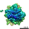 1067MC M: map data used to model this data C: citing same article ( |
|---|---|
| Similar structure data |


-Links
 PDBj
PDBj


- Assembly
| Deposited unit | 
|
|---|---|
| 1 |
|
-Components
-RNA chain , 3 types, 3 molecules AAB3B4
| #1: RNA chain | Mass: 489331.719 Da / Num. of mol.: 1 / Source method: isolated from a natural source Details: modelled as analogous molecule of T. thermophilus taken from PDB entry 1FJF Source: (natural) |
|---|---|
| #18: RNA chain | Mass: 971012.312 Da / Num. of mol.: 1 / Source method: isolated from a natural source Details: REPRESENTED BY THE ANALOGOUS MOLECULE OF H. MARISMORTUI TAKEN FROM PDB ENTRY 1FFK AND SUPPLEMENTED WITH SEQUENCES FROM PDB ENTRIES 1MMS, 1MZP AND 1GIY Source: (natural) |
| #19: RNA chain | Mass: 40298.008 Da / Num. of mol.: 1 / Source method: isolated from a natural source Details: REPRESENTED BY THE ANALOGOUS MOLECULE OF H. MARISMORTUI TAKEN FROM PDB ENTRY 1FFK Source: (natural) |
-Protein , 1 types, 1 molecules AT
| #2: Protein | Mass: 93407.125 Da / Num. of mol.: 1 / Source method: isolated from a natural source / Source: (natural) |
|---|
-40S ribosomal protein ... , 15 types, 15 molecules ABACADAEAGAHAIAJAKALAMANAOAQAS
| #3: Protein | Mass: 20355.412 Da / Num. of mol.: 1 / Source method: isolated from a natural source / Source: (natural) |
|---|---|
| #4: Protein | Mass: 21321.918 Da / Num. of mol.: 1 / Source method: isolated from a natural source / Source: (natural) |
| #5: Protein | Mass: 20363.400 Da / Num. of mol.: 1 / Source method: isolated from a natural source / Source: (natural) |
| #6: Protein | Mass: 15639.166 Da / Num. of mol.: 1 / Source method: isolated from a natural source / Source: (natural) |
| #7: Protein | Mass: 16577.156 Da / Num. of mol.: 1 / Source method: isolated from a natural source / Source: (natural) |
| #8: Protein | Mass: 14518.867 Da / Num. of mol.: 1 / Source method: isolated from a natural source / Source: (natural) |
| #9: Protein | Mass: 15746.292 Da / Num. of mol.: 1 / Source method: isolated from a natural source / Source: (natural) |
| #10: Protein | Mass: 11352.213 Da / Num. of mol.: 1 / Source method: isolated from a natural source / Source: (natural) |
| #11: Protein | Mass: 14459.474 Da / Num. of mol.: 1 / Source method: isolated from a natural source / Source: (natural) |
| #12: Protein | Mass: 12879.160 Da / Num. of mol.: 1 / Source method: isolated from a natural source / Source: (natural) |
| #13: Protein | Mass: 15485.841 Da / Num. of mol.: 1 / Source method: isolated from a natural source / Source: (natural) |
| #14: Protein/peptide | Mass: 4328.041 Da / Num. of mol.: 1 / Source method: isolated from a natural source / Source: (natural) |
| #15: Protein | Mass: 7841.351 Da / Num. of mol.: 1 / Source method: isolated from a natural source / Source: (natural) |
| #16: Protein | Mass: 8777.438 Da / Num. of mol.: 1 / Source method: isolated from a natural source / Source: (natural) |
| #17: Protein | Mass: 8901.519 Da / Num. of mol.: 1 / Source method: isolated from a natural source / Source: (natural) |
+60S ribosomal protein ... , 28 types, 28 molecules BABBBCBDBEBFBGBHBIBJBKBLBMBNBOBPBQBRBSBTBUBVB0BWBXBYBZB9
-Experimental details
-Experiment
| Experiment | Method: ELECTRON MICROSCOPY |
|---|---|
| EM experiment | Aggregation state: PARTICLE / 3D reconstruction method: single particle reconstruction |
- Sample preparation
| Component |
|
|---|