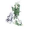
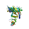
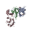
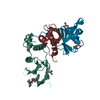
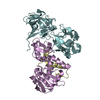
Text: This structure was determined using the XRD structures of plastocyanin and cytochrome f, docked on the basis of intermolecular pseudocontact shifts and chemical shift perturbations due to binding.
-
試料調製
詳細
Solution-ID
内容
溶媒系
1
0.5 mM Cd-substituted poplar plastocyanin + 0.35 mM soluble fragment of turnip cytochrome f in oxidized state in 10 mM sodium phosphate pH 6.0
94% H2O, 6% D2O
2
0.5 mM Cd-substituted poplar plastocyanin + 0.35 mM soluble fragment of turnip cytochrome f in reduced state in 10 mM sodium phosphate pH 6.0 + 1 mM sodium ascorbate
94% H2O, 6% D2O
試料状態
Conditions-ID
イオン強度
pH
圧 (kPa)
温度 (K)
1
10mM
6
1atm
303K
2
11mM
6
1atm
303K
-
NMR測定
放射
プロトコル: SINGLE WAVELENGTH / 単色(M)・ラウエ(L): M
放射波長
相対比: 1
NMRスペクトロメーター
タイプ: Bruker DMX / 製造業者: Bruker / モデル: DMX / 磁場強度: 600 MHz
-
解析
NMR software
名称
バージョン
開発者
分類
Azara
2.7
W. Boucher
解析
ANSIG
Ansig-for-Windows 1.02
P. Kraulis, M. Helgstrand
データ解析
X-PLOR
XPLOR-NIH 2.9.1
構造決定
X-PLOR
XPLOR-NIH 2.9.1
精密化
精密化
手法: Rigid body docking using intermolecular pseudocontact shifts restraints, chemical shift perturbation restraints, electrostatic restraints. ソフトェア番号: 1 詳細: The structures are based on 38 chemical shift perturbation restraints, 42 pseudocontact shifts restraints, 42 pseudocontact angle restraints, 120 minimal distance restraints and 4 ...詳細: The structures are based on 38 chemical shift perturbation restraints, 42 pseudocontact shifts restraints, 42 pseudocontact angle restraints, 120 minimal distance restraints and 4 electrostatic restraints. Backbone RMSD (in Angstrom) with the mean for each model: 1: 1.28, 2: 2.26, 3: 1.29, 4: 3.12, 5: 1.52, 6: 1.49, 7: 3.68, 8: 2.68, 9: 1.58, 10: 3.43, average=2.23+/-0.88
代表構造
選択基準: closest to the average
NMRアンサンブル
コンフォーマー選択の基準: structures with the least restraint violations 計算したコンフォーマーの数: 104 / 登録したコンフォーマーの数: 10
 ムービー
ムービー コントローラー


 データを開く
 基本情報
基本情報 要素
要素 キーワード
キーワード 機能・相同性情報
 データ登録者
 引用
引用 構造の表示
構造の表示 ダウンロードとリンク
 その他のダウンロード


 PDBj
PDBj


 集合体
集合体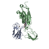


 分子量: 63.546 Da / 分子数: 1 / 由来タイプ: 合成 / 式: Cu
 分子量: 618.503 Da / 分子数: 1 / 由来タイプ: 合成 / 式: C34H34FeN4O4
 HSQC
HSQC 試料調製
試料調製 解析
解析