 Movie
Movie Controller

[English] 日本語
 Yorodumi
- PDB-5pcy: CRYSTAL STRUCTURE ANALYSES OF REDUCED (CUI) POPLAR PLASTOCYANIN A... -
+ Open data

- Basic information
| Entry | Database: PDB / ID: 5pcy | ||||||
|---|---|---|---|---|---|---|---|
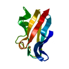
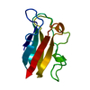
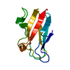
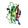
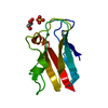
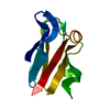
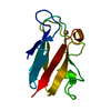
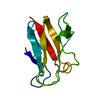
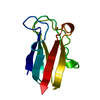
| Title | CRYSTAL STRUCTURE ANALYSES OF REDUCED (CUI) POPLAR PLASTOCYANIN AT SIX PH VALUES | ||||||
 Components Components | PLASTOCYANIN | ||||||
 Keywords Keywords | ELECTRON TRANSPORT PROTEIN(CUPROPROTEIN) | ||||||
| Function / homology |  Function and homology information Function and homology informationchloroplast thylakoid lumen / chloroplast thylakoid membrane / electron transfer activity / copper ion binding Similarity search - Function | ||||||
| Biological species |  | ||||||
| Method |  X-RAY DIFFRACTION / Resolution: 1.8 Å X-RAY DIFFRACTION / Resolution: 1.8 Å | ||||||
 Authors Authors | Guss, J.M. / Freeman, H.C. | ||||||
 Citation Citation | Journal: J.Mol.Biol. / Year: 1986 Title: Crystal structure analyses of reduced (CuI) poplar plastocyanin at six pH values. Authors: Guss, J.M. / Harrowell, P.R. / Murata, M. / Norris, V.A. / Freeman, H.C. #1: Journal: J.Biol.Chem. / Year: 1986Title: The Crystal Structure of Mercury-Substituted Poplar Plastocyanin at 1.9-Angstroms Resolution Authors: Church, W.B. / Guss, J.M. / Potter, J.J. / Freeman, H.C. #2: Journal: J.Biol.Chem. / Year: 1984Title: The Crystal Structure of Poplar Apoplastocyanin at 1.8-Angstroms Resolution. The Geometry of the Copper-Binding Site is Created by the Polypeptide Authors: Garrett, T.P.J. / Clingeleffer, D.J. / Guss, J.M. / Rogers, S.J. / Freeman, H.C. #3: Journal: J.Mol.Biol. / Year: 1983Title: Structure of Oxidized Poplar Plastocyanin at 1.6 Angstroms Resolution Authors: Guss, J.M. / Freeman, H.C. #4: Journal: Nature / Year: 1978Title: X-Ray Crystal Structure Analysis of Plastocyanin at 2.7 Angstroms Resolution Authors: Colman, P.M. / Freeman, H.C. / Guss, J.M. / Murata, M. / Norris, V.A. / Ramshaw, J.A.M. / Venkatappa, M.P. | ||||||
| History |
|
- Structure visualization
| Structure viewer | Molecule: MolmilJmol/JSmol |
|---|
- Downloads & links
-Download
| PDBx/mmCIF format | 5pcy.cif.gz | 30.4 KB | Display | PDBx/mmCIF format |
|---|---|---|---|---|
| PDB format | pdb5pcy.ent.gz | 20.1 KB | Display | PDB format |
| PDBx/mmJSON format | 5pcy.json.gz | Tree view | PDBx/mmJSON format | |
| Others |  Other downloads Other downloads |
-Validation report
| Arichive directory | https://data.pdbj.org/pub/pdb/validation_reports/pc/5pcyftp://data.pdbj.org/pub/pdb/validation_reports/pc/5pcy | HTTPS FTP |
|---|
-Related structure data


-Links
 PDBj
PDBj- Assembly
| Deposited unit | 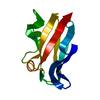
| ||||||||
|---|---|---|---|---|---|---|---|---|---|
| 1 |
| ||||||||
| Unit cell |
| ||||||||
| Atom site foot note | 1: RESIDUES 16 AND 36 ARE CIS-PROLINES. / 2: SEE REMARK 4. |
-Components
| #1: Protein | Mass: 10493.607 Da / Num. of mol.: 1 Source method: isolated from a genetically manipulated source Source: (gene. exp.) |
|---|---|
| #2: Chemical | ChemComp-CU /   Mass: 63.546 Da / Num. of mol.: 1 / Source method: obtained synthetically / Formula: Cu Mass: 63.546 Da / Num. of mol.: 1 / Source method: obtained synthetically / Formula: Cu |
| #3: Water | ChemComp-HOH /  Mass: 18.015 Da / Num. of mol.: 44 / Source method: isolated from a natural source / Formula: H2O Mass: 18.015 Da / Num. of mol.: 44 / Source method: isolated from a natural source / Formula: H2O |
-Experimental details
-Experiment
| Experiment | Method: X-RAY DIFFRACTION |
|---|
- Sample preparation
| Crystal | Density Matthews: 1.92 Å3/Da / Density % sol: 35.93 % | ||||||||||||||||||||
|---|---|---|---|---|---|---|---|---|---|---|---|---|---|---|---|---|---|---|---|---|---|
| Crystal grow | *PLUS pH: 6 / Method: vapor diffusion / Details: Chapman, G.V. (1977). J. Mol. Biol., 110, 187. | ||||||||||||||||||||
| Components of the solutions | *PLUS
|
-Data collection
| Reflection | *PLUS Highest resolution: 1.8 Å / Lowest resolution: 7 Å / Num. obs: 6971 / Num. measured all: 9478 / Rmerge(I) obs: 0.078 |
|---|
- Processing
| Software | Name: PROLSQ / Classification: refinement | ||||||||||||
|---|---|---|---|---|---|---|---|---|---|---|---|---|---|
| Refinement | Rfactor obs: 0.16 / Highest resolution: 1.8 Å Details: THE SHORT CONTACT BETWEEN CE1 HIS 87 AND O PRO 36 (3.4 ANGSTROMS AT PH 7.8, 3.3 ANGSTROMS AT PH 7.0) IS REPLACED BY A WEAK HYDROGEN BOND (3.1 ANGSTROMS) BETWEEN NE2 HIS 87 AND O PRO 36 AFTER ...Details: THE SHORT CONTACT BETWEEN CE1 HIS 87 AND O PRO 36 (3.4 ANGSTROMS AT PH 7.8, 3.3 ANGSTROMS AT PH 7.0) IS REPLACED BY A WEAK HYDROGEN BOND (3.1 ANGSTROMS) BETWEEN NE2 HIS 87 AND O PRO 36 AFTER ROTATION OF THE HIS 87 SIDE CHAIN AT PH 3.8 (SEE *JRNL* REFERENCE ABOVE). | ||||||||||||
| Refinement step | Cycle: LAST / Highest resolution: 1.8 Å
| ||||||||||||
| Refinement | *PLUS Lowest resolution: 7 Å / Num. reflection obs: 6900 / Rfactor obs: 0.16 | ||||||||||||
| Solvent computation | *PLUS | ||||||||||||
| Displacement parameters | *PLUS |
