 Movie
Movie Controller


+ Open data

- Basic information
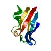
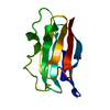
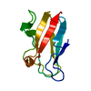
| Entry | Database: PDB / ID: 1byp | ||||||
|---|---|---|---|---|---|---|---|
| Title | E43K,D44K DOUBLE MUTANT PLASTOCYANIN FROM SILENE | ||||||
 Components Components | PROTEIN (PLASTOCYANIN) | ||||||
 Keywords Keywords | ELECTRON TRANSPORT / ELECTRON TRANSFER / PHOTOSYNTHESIS / ACIDIC PATCH / DOUBLE MUTANT | ||||||
| Function / homology |  Function and homology information Function and homology informationchloroplast thylakoid lumen / chloroplast thylakoid membrane / electron transfer activity / copper ion binding Similarity search - Function | ||||||
| Biological species |  Silene latifolia subsp. alba (white campion) Silene latifolia subsp. alba (white campion) | ||||||
| Method |  X-RAY DIFFRACTION / MOLECULAR REPLACEMENT / Resolution: 1.75 Å X-RAY DIFFRACTION / MOLECULAR REPLACEMENT / Resolution: 1.75 Å | ||||||
 Authors Authors | Sugawara, H. / Inoue, T. / Li, C. / Gotowda, M. / Hibino, T. / Takabe, T. / Kai, Y. | ||||||
 Citation Citation | Journal: J.Biochem.(Tokyo) / Year: 1999 Title: Crystal structures of wild-type and mutant plastocyanins from a higher plant, Silene. Authors: Sugawara, H. / Inoue, T. / Li, C. / Gotowda, M. / Hibino, T. / Takabe, T. / Kai, Y. #1: Journal: Acta Crystallogr.,Sect.D / Year: 1997Title: Crystallization and Preliminary X-Ray Studies of Plastocyanin from Silene Expressed in E. Coli Authors: Li, C. / Inoue, T. / Gotowda, M. / Hamada, K. / Nishio, N. / Hibino, T. / Takabe, T. / Kai, Y. #2: Journal: Acta Crystallogr., Sect.B / Year: 1992Title: Accuracy and Precision in Protein Structure Analysis: Restrained Least-Squares Refinement of the Structure of Poplar Plastocyanin at 1.33 A Resolution Authors: Guss, J.M. / Bartunik, H.D. / Freeman, H.C. | ||||||
| History |
|
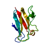
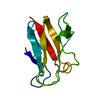
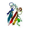
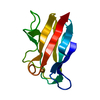
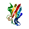
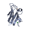
- Structure visualization
| Structure viewer | Molecule: MolmilJmol/JSmol |
|---|
- Downloads & links
-Download
| PDBx/mmCIF format | 1byp.cif.gz | 32.1 KB | Display | PDBx/mmCIF format |
|---|---|---|---|---|
| PDB format | pdb1byp.ent.gz | 21.4 KB | Display | PDB format |
| PDBx/mmJSON format | 1byp.json.gz | Tree view | PDBx/mmJSON format | |
| Others |  Other downloads Other downloads |
-Validation report
| Arichive directory | https://data.pdbj.org/pub/pdb/validation_reports/by/1bypftp://data.pdbj.org/pub/pdb/validation_reports/by/1byp | HTTPS FTP |
|---|
-Related structure data
| Related structure data | 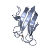 1byoC  1pcy C: citing same article ( S: Starting model for refinement |
|---|---|
| Similar structure data |

-Links
 PDBj
PDBj- Assembly
| Deposited unit | 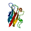
| ||||||||
|---|---|---|---|---|---|---|---|---|---|
| 1 |
| ||||||||
| Unit cell |
|
-Components
| #1: Protein | Mass: 10415.667 Da / Num. of mol.: 1 / Mutation: E43K, D44K Source method: isolated from a genetically manipulated source Source: (gene. exp.) Silene latifolia subsp. alba (white campion)Species: Silene latifolia / Strain: subsp. alba / Organelle: CHLOROPLAST / Production host:  |
|---|---|
| #2: Chemical | ChemComp-CU /   Mass: 63.546 Da / Num. of mol.: 1 / Source method: obtained synthetically / Formula: Cu Mass: 63.546 Da / Num. of mol.: 1 / Source method: obtained synthetically / Formula: Cu |
| #3: Water | ChemComp-HOH /  Mass: 18.015 Da / Num. of mol.: 73 / Source method: isolated from a natural source / Formula: H2O Mass: 18.015 Da / Num. of mol.: 73 / Source method: isolated from a natural source / Formula: H2O |
-Experimental details
-Experiment
| Experiment | Method: X-RAY DIFFRACTION / Number of used crystals: 1 |
|---|
- Sample preparation
| Crystal | Density Matthews: 2.54 Å3/Da / Density % sol: 52 % | ||||||||||||||||||||
|---|---|---|---|---|---|---|---|---|---|---|---|---|---|---|---|---|---|---|---|---|---|
| Crystal grow | pH: 7 Details: 2.8M AMMONIUM SULFATE, 100MM POTASSIUM PHOSPHATE, pH 7.0 | ||||||||||||||||||||
| Crystal grow | *PLUS Method: vapor diffusion, hanging drop | ||||||||||||||||||||
| Components of the solutions | *PLUS
|
-Data collection
| Diffraction | Mean temperature: 300 K |
|---|---|
| Diffraction source | Source: ROTATING ANODE / Type: RIGAKU RU300 / Wavelength: 1.5418 |
| Detector | Type: RIGAKU / Detector: IMAGE PLATE / Date: Jan 15, 1995 |
| Radiation | Monochromator: NI FILTER / Protocol: SINGLE WAVELENGTH / Monochromatic (M) / Laue (L): M / Scattering type: x-ray |
| Radiation wavelength | Wavelength: 1.5418 Å / Relative weight: 1 |
| Reflection | Resolution: 1.75→30 Å / Num. obs: 10737 / % possible obs: 93.6 % / Observed criterion σ(I): 0 / Redundancy: 4.9 % / Rmerge(I) obs: 0.056 |
| Reflection | *PLUS Num. measured all: 52627 |
- Processing
| Software |
| ||||||||||||||||||||||||||||||||||||||||||||||||||||||||||||||||||||||||||||||||||||
|---|---|---|---|---|---|---|---|---|---|---|---|---|---|---|---|---|---|---|---|---|---|---|---|---|---|---|---|---|---|---|---|---|---|---|---|---|---|---|---|---|---|---|---|---|---|---|---|---|---|---|---|---|---|---|---|---|---|---|---|---|---|---|---|---|---|---|---|---|---|---|---|---|---|---|---|---|---|---|---|---|---|---|---|---|---|
| Refinement | Method to determine structure: MOLECULAR REPLACEMENT Starting model: PDB ENTRY 1PCY 1pcy Resolution: 1.75→20 Å / Cross valid method: THROUGHOUT / σ(F): 0
| ||||||||||||||||||||||||||||||||||||||||||||||||||||||||||||||||||||||||||||||||||||
| Refinement step | Cycle: LAST / Resolution: 1.75→20 Å
| ||||||||||||||||||||||||||||||||||||||||||||||||||||||||||||||||||||||||||||||||||||
| Refine LS restraints |
| ||||||||||||||||||||||||||||||||||||||||||||||||||||||||||||||||||||||||||||||||||||
| Software | *PLUS Name: REFMAC / Classification: refinement | ||||||||||||||||||||||||||||||||||||||||||||||||||||||||||||||||||||||||||||||||||||
| Refinement | *PLUS Lowest resolution: 20 Å / σ(F): 0 / % reflection Rfree: 5 % / Rfactor obs: 0.18 | ||||||||||||||||||||||||||||||||||||||||||||||||||||||||||||||||||||||||||||||||||||
| Solvent computation | *PLUS | ||||||||||||||||||||||||||||||||||||||||||||||||||||||||||||||||||||||||||||||||||||
| Displacement parameters | *PLUS |
